Background
Hepatocellular Carcinoma (HCC) is one of the most common malignancies in adults [1]. Management of patients with HCC depends on the stage of their tumor. Portal Vein Tumor Thrombosis (PVTT) is a common condition in patients with advanced HCC [2]. PVTT may cause serious complications such as tumor spread, deterioration of liver function and portal vein hypertension, leading to intractable ascites, variceal rupture, hepatic encephalopathy or death [3].
The Child–Pugh classification system has been widely used to predict the risk of death and complications in patients with cirrhosis. The Child–Pugh class B liver function, extensive venous involvement and elevated tumor markers have been identified as poor prognostic factors in RT for HCC [4].
Decisions about the optimal treatment for HCC are complex because many therapeutic options need to be considered based on the tumor burden, underlying hepatic function, patient performance status, and availability or applicability of specific treatment modalities [5]. For example, not all patients with early-stage HCC can undergo curative treatments, such as hepatic resection, Radiofrequency Ablation (RFA), or Liver Transplantation (LT), because of various clinical conditions [6]. Moreover, tumor recurrences are frequently observed after initial treatment; however, there are no clear recommendations or guidelines for the management of recurrent HCC. Due to this complexity of treatment decisions, a multidisciplinary approach is recommended to maximize therapeutic efficacy [7].
Historically, Radiation Therapy (RT) for liver tumor was limited because liver was known to be a radiosensitive organ. Yet with advances in imaging, treatment planning, and treatment delivery, now we could try to deliver tumoricidal doses to target areas without incurring significant side effects. Partial liver irradiation has shown some promising results in patients with unresectable HCC; promising outcomes were also observed in patients with PVT who were treated with RT [8,9].
Methods study design and conduct
This phase III nonrandomized controlled study was conducted at the Clinical Oncology Department of Suez Canal University Hospital in Ismailia governorate, Egypt, from June 2022 to May 2024. The aim of the study was to assess the tumor response rate by radiological and laboratory response in addition to overall survival and progression free survival by using 3D Conformal radiotherapy directed to liver mass in Hepatocellular carcinoma patients. The study protocol was approved by the Research Ethics Committee at the institute, and all patients provided written informed consent before participating in the study. Patients had the right to withdraw from the study at any time. The authors take full responsibility for the accuracy, confidentiality of data, and adherence to the study protocol.
Patients
The sample size was calculated as 14 patients in each group.
Group A: The experimental arm received external beam radiotherapy localized to the hepatic mass. Group B: The control arm received the standard treatment which was supportive treatment.
Eligible patients were pathological or radiological proven HCC patients, Patients with unresectable disease, or who were medically inoperable due to comorbidity and unfit for surgery, patients who had comorbidities interfere with usage of Targeted and Immunotherapy and a performance status of 0-2 according to the Eastern Cooperative Oncology Group performance scale and fall within the Child-Pugh A or B classification. Patients with Tumor size totally do not exceed 10 cm.
We excluded patients who had small sized lesion eligible for Surgery, or who received definitive targeted therapy and Immunotherapy, or Patients with Performance Status >2.
Randomization and masking
Patients were nonrandomly allocated by the investigators and open labelled, the 28 patients divided into 2 groups, each group consisting of 14 patients.
Intervention
All patients were divided into two groups. Patients were randomly assigned to either radiotherapy or control arms. Each group consists of 14 patients.
Group A: Experimental arm in which we received palliative conformal three-dimensional radiotherapy on Liver mass. Group B: Control arm received best supportive care (palliative chemotherapy, palliative radiotherapy or supportive treatment).
Technique of external beam radiotherapy
CT Simulation and patient set up: CT simulation is performed with 3 mm slice thickness. Bowel prepared night prior by rectal voiding and low residue diet after treatment is advised to avoid diarrhea. IV contrast may aid in delineation of Hepatic lesion. Planning CT is obtained in treatment position, Patient is positioned supine and fixed with arms under his head. Skin reference tattoos are placed to prevent lateral rotation. Radio-opaque markers are placed on skin to locate the tattoos on CT scans. Scan from chest to pelvis to allow for delineation of Heart, Spinal cord, both Kidneys and relevant small bowel areas adjacent to high dose distributions. Image guided verification and adjustment will be done 3 times in the first week then one weekly for the remaining course.
Target volume delineation and treatment planning: GTV: gross tumor shown on the on the arterial-phase CT scan.
CTV: An additional margin of 1 cm around the GTV, confined to the liver to form a Clinical Target Volume (CTV) to account for microscopic extensions and internal movement as (ITV).
PTV: expanded from CTV to three-dimensional, typically with margins of 5 mm, accounting for Set up error. Planning systems: (Monaco, Elekta, Stockholm, Sweden; Eclipse, Varian, Palo Alto, USA).
Dose prescription: 50 Gy over 25 Fractions, 2 Gy per Fraction.
For the assessment of tumor response rate by radiological and laboratory response pretreatment and after 1 month of finishing the management, we classified the tumor response rate into 3 categories (below 25%, 25-50% and above 50%), and overall survival and progression free survival by using the Kaplan Meier curves.
Table 1: Patient and tumor characteristics, laboratory parameters, performance status, underlying liver disease and child pugh score.
| |
Control |
Radiotherapy |
P-Value |
| N:14 |
% |
N:14 |
% |
| Age |
|
Median 69(53-85) |
Median 63(52-74) |
0.296 |
| Gender |
Male |
11 |
78.6 |
10 |
71.4 |
0.676 |
| Female |
3 |
21.4 |
4 |
28.6 |
| Comorbidities |
DM |
1 |
7.1 |
3 |
21.4 |
0.306 |
| HTN |
6 |
42.9 |
2 |
14.3 |
| Both |
1 |
7.1 |
3 |
21.4 |
| No |
6 |
42.9 |
6 |
42.9 |
| Pathologicalproven |
Yes |
10 |
71.4 |
9 |
64.3 |
0.699 |
| No |
4 |
28.6 |
5 |
35.7 |
| Stage |
T4N1 |
7 |
50 |
8 |
57.1 |
0.568 |
| T4N0 |
5 |
35.7 |
2 |
14.3 |
| T3N1 |
2 |
14.3 |
2 |
14.3 |
| T3N0 |
0 |
0 |
2 |
14.3 |
| Size |
<5 cm |
2 |
14.3 |
0 |
0 |
0.153 |
| >5 cm |
12 |
85.7 |
14 |
100 |
| Site |
One lobe |
12 |
85.7 |
13 |
92.9 |
0.237 |
| Both lobes |
2 |
14.3 |
1 |
7.1 |
| PVT |
Yes |
12 |
85.7 |
13 |
92.9 |
0.558 |
| No |
2 |
14.3 |
1 |
7.1 |
| Child Pughscale |
A |
6 |
42.9 |
5 |
35.7 |
|
| B |
8 |
57.1 |
9 |
64.3 |
| ECOG |
0 |
2 |
14.3 |
2 |
14.3 |
0.793 |
| 1 |
6 |
42.9 |
7 |
50 |
| 2 |
6 |
42.9 |
5 |
35.7 |
| AFP |
|
Range (14-2955) |
Range (0.9-200000) |
0.355 |
| Liverenzymes level(ALT, AST) |
|
Range (35-210) |
Range (25-190) |
| INR |
Normal |
9 |
64.3 |
6 |
42.9 |
0.272 |
| High |
5 |
35.7 |
8 |
57.1 |
| Serum bilirubin |
Normal |
12 |
85.7 |
5 |
35.7 |
0.005* |
| High |
2 |
14.3 |
9 |
64.3 |
Data collection was systematically conducted, and all data were anonymized for analysis. The statistical analysis was performed using SPSS version 25.
Results
We enrolled 28 patients with Locally advanced Hepatocellular Carcinoma who were unfit for resection, radiofrequency ablation, and unavailability of targeted therapy or Immunotherapy attending Department of Clinical Oncology in Suez Canal University Hospital at the time from June 2022 till May 2024.
Patients were non-randomly distributed between two groups:
Group A: 14 patients received external beam radiotherapy localized to the hepatic mass.
Group B: Another 14 patients received the standard treatment which was best supportive care.
Table 2: Treatment outcomes and response rate in control arm versus patient treated with radiotherapy.
| |
Control |
Radiotherapygroup |
P-Value |
| N:14 |
% |
N:14 |
% |
| Tumor size |
Regression |
0 |
0 |
13 |
92.9 |
<0.001* |
| Partial |
0 |
0 |
13 |
92.9 |
| Complete |
0 |
0 |
0 |
0 |
| Stationary |
9 |
64.3 |
1 |
7.1 |
| Progression |
5 |
35.7 |
0 |
0 |
The study’s findings, summarized in several tables and figures, provide insights into the treatment’s efficacy. Patient characteristics, detailed in (Table 1). A total of 28 patients were enrolled, the median age was 69 years old in the control group and 63 years old in the radiotherapy group. Most patients were male (21 out of 28). Table 1 also showed the tumor characteristics, performance status profile according to ECOG score, and Child Pugh scale before starting the management. All patients were within the Child-Pugh A or B stage, indicative of less severe liver disease. The laboratory investigations regarding AFP, Liver enzymes (ALT-AST), serum bilirubin and INR, with elevated serum bilirubin in favor of radiotherapy arm due to intra hepatic biliary obstruction in 9 patients.
Table 3: Treatment outcomes regarding laboratory parameters in control arm versus patient treated with radiotherapy.
| |
Control |
Radiotherapy |
P-Value |
| N:14 |
% |
N:14 |
% |
| AFP |
Improved |
8 |
57.1 |
13 |
92.9 |
0.082 |
| Stationary |
2 |
14.3 |
0 |
0 |
| Deteriorate |
4 |
28.6 |
1 |
7.1 |
| Liver enzymes |
Improved |
6 |
42.8 |
3 |
21.4 |
|
| Stationary |
4 |
28.6 |
10 |
71.4 |
| Deteriorate |
4 |
28.6 |
1 |
7.1 |
| S.bilirubin |
Improved |
2 |
14.3 |
8 |
57.1 |
0.024* |
| Stationary |
12 |
85.7 |
5 |
35.7 |
| Deteriorate |
0 |
0 |
1 |
7.1 |
Table 4: Multivariate analysis of factors affects radio morphological response in patients treated with radiotherapy.
| Factor |
Odds (95% confidence index |
P-value |
| Stage |
0.700 (0.88-5.564) |
0.736 |
| Child-Pugh score |
0.000 (0.000-1.806) |
0.066 |
| ECOG |
0.009 (0.000-4.595) |
0.14 |
| AFP |
0.998 (0.996-1.000) |
0.112 |
| Liver enzymes |
0.000 (0.000-5.084) |
0.103 |
| INR |
11.797(0.027-5081.722) |
0.425 |
| RT dose |
6.981(0.361-135.143) |
0.199 |
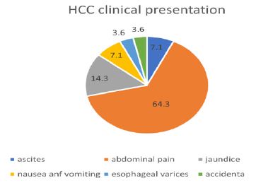
Figure 1 showed a pie chart of clinical presentation of all HCC patients involved in our study, 64.3% of the patients complained of abdominal pain as the initial presentation at diagnosis, while 14.3% presented with obstructive jaundice and 7.1% complained of nausea and vomiting and another 7.1% complained of ascites. 3.6% of the patients discovered accidentally that they had HCC (P-value <0.001).
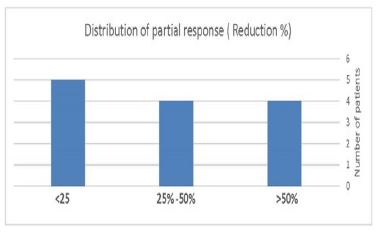
Regarding (Table 2 and Figure 2), they showed that there were 13 patients of the 14 of radiotherapy group who achieved partial regression in the size of tumor, 5 patients of them achieved <25% reduction in size, while 4 patients of them achieved 25-50% reduction in tumor size and another 4 patients achieved >50% reduction in tumor size. Only 1 patient of the radiotherapy group was stationary course in tumor size.
In the control group, 9 patients who achieved stationary course in tumor size, while the rest of the group achieved progressive course.
Table 3 showed improvement in the laboratory investigations in the tumor marker and especially in the level of serum bilirubin in the radiotherapy group, as we started the treatment with 9 patients complained of elevated serum bilirubin and after the treatment 8 patients achieved marked improvement with significant (P-value 0.024). (Table 4) showed that there was a statistical significance of Child-Pugh score with (P-value 0.066) and this means that Child-Pugh score was the most factor that affects the radiotherapy treatment.
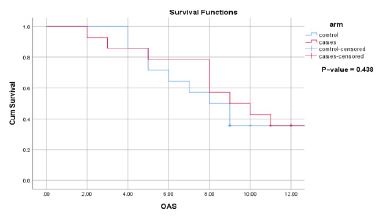
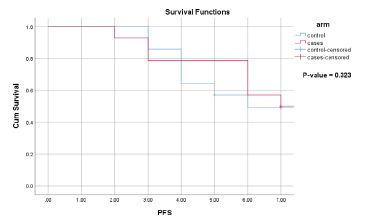
(Figures 3 and 4) showed the difference of PFS between patients treated with radiotherapy and control arm was statistically insignificant (p-value=0.323). The PFS median was 6.5 months, and The OAS median was 9 months (p-value=0.438).
Discussion
Three-dimensional radiotherapy was administered to patients with locally advanced Hepatocellular Carcinoma (HCC) who were unfit for resection or radiofrequency ablation and unavailability targeted therapy or Immunotherapy, resulting in a clinically significant improvement in Tumor size and laboratory investigations after one month of treatment. There was partial regression of the tumor size in 13 patients with a percentage of 92.9% with (P-value 0.001) in comparison with control Group and only there was 1 patient who was stationary, and no one reached complete response [10,11].
In our study, we did a multivariate analysis of multiple factors such Stage, Child-Pugh score, ECOG, AFP, pain score, Liver enzymes, INR, RT dose, that affects radio morphological response in patients treated with radiotherapy and showed that Child-Pugh score with (P-value 0.066) and this means that Child-Pugh score is the factor that affects the radiotherapy treatment group [12,13].
The Progression free survival between patients treated with radiotherapy and control group was statistically insignificant (p-value=0.323). The PFS median was 6.5 months, While the overall survival median was 9 months with (p-value=0.438) [14].
Conclusion
In conclusion, external beam radiotherapy is considered effective treatment in management of Locally advanced HCC causing improving the tumor size and relatively improved laboratory investigations especially in the patients presented with obstructive jaundice because of obstruction of the Common Bile duct or the intrahepatic biliary radicals. But using 3DCRT didn’t improve the OS and PFS in comparison with patients treated with radiotherapy and the patients treated with best supportive care.
References
- El-Serag HB. Hepatocellular carcinoma. N Engl J Med. 2011; 365: 1118–27.
- Pirisi M, Avellini C, Fabris C, et al. Portal vein thrombosis in hepatocellular carcinoma: age and sex distribution in an autopsy study. J Cancer Res Clin Oncol. 1998; 124: 397–400.
- Takizawa D, Kakizaki S, Sohara N, et al. Hepatocellular carcinoma with portal vein tumor thrombosis: clinical characteristics, prognosis, and patient survival analysis. Dig Dis Sci. 2007; 52: 3290–5.
- Roudot-Thoraval F. Epidemiology of hepatitis C virus infection. Clin Res Hepatol Gastroenterol. 2021; 45: 101596.
- Kamarajah SK, Frankel TL, Sonnenday C, et al. Critical evaluation of the American Joint Commission on Cancer (AJCC) 8th edition staging system for patients with Hepatocellular Carcinoma (HCC): a Surveillance, Epidemiology, End Results (SEER) analysis. J Surg Oncol. 2018; 117: 644–50.
- Chang W, Lee JM, Lee DH, et al. Comparison of switching bipolar ablation with multiple cooled wet electrodes and switching monopolar ablation with separable clustered electrode in treatment of small hepatocellular carcinoma: a randomized controlled trial. PLoS One. 2018; 13: e0192173.
- Yopp AC, Mansour JC, Beg MS, et al. Establishment of a multidisciplinary hepatocellular carcinoma clinic is associated with improved clinical outcome. Ann Surg Oncol. 2014; 21: 1287–95.
- Akinori Asagi, Hiroshi Ishii, Kazuhiro Uesugi, et al. Three-dimensional conformal radiotherapy (3D- CRT) for Macroscopic Vascular Invasion (MVI) of advanced hepatocellular carcinoma (HCC). 2016: 34.
- Smith Apisarnthanarax, Aisling Barry, Minsong Cao, et al.. External Beam Radiation Therapy for Primary Liver Cancers: An ASTRO Clinical Practice Guideline. 2021; 12: P28-51.
- Iwamoto H, Nomiyama M, Niizeki T, et al. Dose and Location of Irradiation Determine Survival for Patients with Hepatocellular Carcinoma with Macrovascular Invasion in External Beam Radiation Therapy. Oncology. 2019; 96: 192-199.
- Eiichiro Okazaki, Akira Yamamoto, Norifumi Nishida, et al. Three-dimensional conformal radiotherapy for locally advanced hepatocellular carcinoma with portal vein tumour thrombosis: evaluating effectiveness of the model for end-stage liver disease (MELD) score compared with the Child–Pugh classification, British Journal of Radiology 2016; 89: 20150945.
- Bae Bong Kyung, Kim Jae-Chul. The response of thrombosis in the portal vein or hepatic vein in hepatocellular carcinoma to radiation therapy. Radiat Oncol J. 2016; 34: 168-176.
- Chai Hong Rim, Dae Sik Yang, Young Je Park, et al. Effectiveness of High-dose Threedimensional Conformal Radiotherapy in Hepatocellular Carcinoma with Portal Vein Thrombosis, Japanese Journal of Clinical Oncology. 2012; 42: 721–729.
- Kouloulias Vassilis, Mosa Eftychia, Georgakopoulos J, et al. Three-Dimensional Conformal Radiotherapy for Hepatocellular Carcinoma in Patients Unfit for Resection, Ablation, or Chemotherapy: A Retrospective Study, The Scientific World Journal. 2013; 2013: 780141.