Introduction
Breast cancer is the most prevalent malignant tumor in women and one of the major contributors to the global burden of disease. It holds the position as the second most frequently prevalent cancer and the fourth leading cause of cancer-related mortality worldwide, with 2.3 million new cases and 666,000 fatalities reported in 2022, according to the latest global cancer statistics [1]. Despite significant and substantial progress has been made in drug development for breast cancer, the effectiveness of treatment is still hampered by metastasis and therapeutic resistance. Taxanes, such as Paclitaxel (PTX) and Docetaxel (DTX), is widely used as a first-line chemotherapy agent in the adjuvant and neoadjuvant treatment of breast cancer, especially in advanced metastatic cancer treatment. However, the resistance to taxanes in breast cancer is a huge obstacle to clinical application, which is closely correlated with adverse reactions, recurrence and metastasis. Meanwhile, the generation of drug resistance is also a major cause of death in patients receiving treatment [2].
For the past several years, the possible significance of the Tumor Microenvironment (TME) in tumor development, metastasis, and treatment has been extensively investigated [3,4]. CAFs are the essential components of the TME and are activated fibroblasts with functional heterogeneity [5]. Cancer-Associated Fibroblasts (CAFs) secrete and release a wide range of active biomolecules, such as cytokines, growth factors, Extracellular Matrix (ECM) components, hormones and proteases, which not only promote tumourigenesis, growth and angiogenesis, but also lead to the production and remodeling of aberrant extracellular matrices, promoting tumor migration, invasion and distant metastasis [6,7]. At the same time, CAFs can develop or enhance chemoresistance through various mechanisms, including induction of EpithelialMesenchymal Transition (EMT), activation of stem cell-related programs, and reprogramming of tumor cell metabolism [8].
Increasing evidence indicates a close link between CAFs and breast cancer. For example, the oncogenic function of CAFs in breast cancer is dependent on STAT3, which acts as a tumor growth promoter by inducing the CAFs secreted proteins ANGPTL4, MMP 13 and STC-1 [9]. In addition, CAFs activated by TGFβ 1 can result in cellular autophagy or overexpression of FAPα, thereby promoting invasion, metastasis, and EMT of breast cancer [10]. Thus, the presence of CAFs is usually significantly associated with poor prognosis in breast cancer [11]. Another explanation for this correlation is that CAFs directly influence the response to treatment and may be involved in the induction of treatment resistance [12]. CD63+ CAFs secrete exosomal miR-22, mediated by SFRS 1, for binding to their targets ER α and PTEN, rendering breast tumor cells resistant to tamoxifen [13]. Given these findings, identifying genes and pathways related to CAFs in breast cancer chemotherapy resistance may facilitate the development of new targets for reducing breast cancer treatment resistance.
Bioinformatics analysis, which advances the development of targeted and individualized medicines, is a useful means for recognizing possible molecular targets for therapy and biomarkers of progression [14-16]. The Cancer Genome Atlas (TCGA) contains information on numerous human malignancies and provides a comprehensive view of similarities, variations and emerging events within neoplasm genealogy [17]. Additionally, the Gene Expression Omnibus (GEO) offers a comprehensive gene expression profile that demonstrates an integrated whole picture of cellular regulation [18]. Using data from the TCGA and GEO databases, this study attempted to find out crucial genes and pathways associated to the regulation of CAFs in chemoresistance in breast cancer.
Materials and methods Data download
Gene expression profiling data of the breast cancer dataset GSE42568 and the breast cancer chemotherapy resistance datasets GSE28784 and GSE99225 were download from the GEO database (https://www.ncbi.nlm.nih.gov/gds/). Both GSE42568 and GSE99225 datasets were obtained from Homo Sapiens, sourced from Breast Tissue, and GSE28784 dataset obtained from Homo Sapiens, sourced from MDA-MB-231 cells. GSE42568 includes 17 normal breast tissue samples and 104 breast cancer tissue samples. GSE99225 includes 4 chemo-sensitive breast tissue samples and 4 chemo-resistant breast cancer tissue samples. GSE28784 includes 3 chemo-sensitive cell lines and 6 chemo-resistant cell lines. All samples were included in this study.
Fibroblast-Related Genes (FRGs) were collected from GeneCards database (https://www.genecards.org/). Using «Fibroblast» as a search term and keeping only «Protein Coding», a total of 19942 FRGs were obtained. At the same time, the top 2000 FRGs were obtained for subsequent analysis in descending order of ‘Relevance Score’.
Clinical information and overall survival data for crucial genes were collected from the TCGA-BRCA at The Cancer Genome Atlas (TCGA) website (https://portal.gdc.cancer.gov/) and the University of California Santa Cruz (UCSC) Xena database (http://xena. ucsc.edu/).
Analysis of differentially expressed genes (DEGs)
According to the grouping of the datasets, the GSE42568 samples were divided into the breast cancer group and the control group, and the GSE28784 and GSE99225 samples were divided into the chemo-sensitive group and the chemo-resistant group. Gene differential expression analysis of each dataset was performed with |log FC|>1.0 and adj. p<0.05 as the threshold, and the results were presented by drawing the volcano plots.
GO and KEGG analysis
Gene Ontology (GO) and Kyoto Encyclopedia of Genes and Genomes (KEGG) analyses were performed were performed to investigate the functional roles of DEGs in Biological Processes (BP), Molecular Functions (MF), and Cellular Components (CC), as well as the associated pathways.
Construction of the PPI network
Protein-Protein Interaction (PPI) networks of candidate genes were constructed using the String database (https://cn.string-db. org/) defining the species as human. Subsequently, the resulting PPI network was imported into Cytoscape software for visualization, and the candidate genes were ranked based on degree value (number of interactions with candidate genes) using the plugin CytoHubba, and the top 20 genes were selected for presentation.
Identification of the intersect genes
The DEGs from breast cancer dataset GSE42568 were intersected with the top 2000 FRGs obtained from GeneCards database to obtain Fibroblast-related Differentially Expressed Genes (FRDEGs) for breast cancer, and a Venn diagram was plotted.
The DEGs from breast cancer chemotherapy resistance dataset GSE99225 were intersected with the top 2000 FRGs obtained from GeneCards database to obtain fibroblast-related chemotherapy resistance genes for breast cancer, and a Venn diagram was plotted.
The crucial genes were obtained by intersecting genes with high degree values in the PPI networks (PPI-top20) with top genes in crucial pathway enrichment analysis (KEGG-top20).
Expression and correlation analysis of the crucial genes
The mRNA expression levels of crucial genes in breast cancer tissues and normal tissues was analyzed based on the TCGA-BRCA dataset, and the results were presented by box-and-line plots. While, the correlations between crucial genes in breast cancer was analyzed and shown by chord plot.
Survival analysis of the crucial genes
The effects of crucial genes on patient prognosis, such as Overall Survival (OS), Disease-Free Survival (DSS) and Progression-Free Interval (PFI), were assessed based on the TCGA-BRCA dataset, and the results were plotted on the Kaplan-Meier (KM) survival curves. The time-dependent Receiver Operating Characteristic (ROC) curves based on the expression of crucial genes and OS were generated, and the Area Under the Curve (AUC) values were calculated to assess diagnostic capacity for breast cancer samples at 1, 3, and 5 years.
Correlation analysis of crucial genes and clinicopathological parameters
Based on the TCGA-BRCA dataset, the correlation between crucial genes and clinical characteristics of breast cancer patients was investigated, including age, menstrual status, anatomical site, pathological TNM stage, expression status of ER, PR, and HER2, and molecular typing.
Immune infiltration analysis
Correlations between crucial genes and major immune cells were analyzed through the TCGA database and represented by a heatmap.
Results
FoxO, TNF, Hippo, Wnt, Rap 1, MAPK, IL-17, ErbB, PI3K-Akt, JAK-STAT signaling pathways are pathways associated with chemoresistance in breast cancer
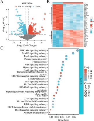
To explore the specific mechanisms affecting chemoresistance in breast cancer, we obtained the breast cancer chemotherapy resistance dataset GSE28784 from the GEO database. The results of differential analysis showed that GSE28784 contained a total of 1041 DEGs, which were demonstrated by volcano and heat maps (Figure 1A-B). KEGG analysis showed that these genes were mainly enriched in 96 pathways, such as FoxO signaling pathway, Th17 cell differentiation, TNF signaling pathway, Hippo signaling pathway, Wnt signaling pathway, Proteoglycans in cancer, Rap1 signaling pathway, EGFR tyrosine kinase inhibitor resistance, MAPK signaling pathway, IL-17 signaling pathway, ErbB signaling pathway, PI3K-Akt signaling pathway, Cellular senescence, Apoptosis, Th1 and Th2 cell differentiation, Focal adhesion, NOD-like receptor signaling pathway, B cell receptor signaling pathway, Transcriptional misregulation in cancer, Signaling pathways regulating pluripotency of stem cells, Platinum drug resistance, JAKSTAT signaling pathway, and Cell cycle (Figure 1C).
PI3K-Akt, EGFR, Ras, Rap 1, MAPK, FoxO, HIF-1, TNF, AMPK, ErbB, PPAR, and PD(L) 1 signaling pathways are pathways associated with CAFs in breast cancer
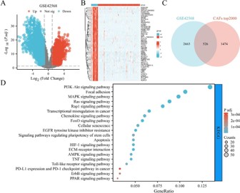
To explore the specific mechanisms of CAFs affecting breast cancer, we obtained the breast cancer dataset GSE42568 from the GEO database. The results of differential analysis showed that GSE42568 contained a total of 3198 DEGs, which were demonstrated by volcano and heat maps (Figure 2A-B). By taking the intersection of DEGs from the breast cancer dataset GSE42568 with the top 2000 FRGs obtained from the GeneCards database, a total of 526 FRDEGs were obtained (Figure 2C). And the signaling pathways in which FRDEGs were mainly enriched were identified using KEGG, and the results showed that FRDEGs were involved in the regulation of 144 pathways, such as Focal adhesion, PI3KAkt signaling pathway, EGFR tyrosine kinase inhibitor resistance, Ras signaling pathway, Rap1 signaling pathway, MAPK signaling pathway, ECM-receptor interaction, FoxO signaling pathway, HIF1 signaling pathway, Transcriptional misregulation in cancer, TNF signaling pathway, Chemokine signaling pathway, Cellular senescence, AMPK signaling pathway, Apoptosis, Toll-like receptor signaling pathway, Signaling pathways regulating pluripotency of stem cells, ErbB signaling pathway, PPAR signaling pathway, PD-L1 expression, and PD-1 checkpoint pathway in cancer (Figure 2D).
Chemoresistance in breast cancer is affected by the CAFs
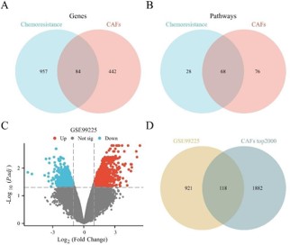
Comparison of the results from analyses of chemoresistant and CAFs revealed 84 shared genes and 68 shared signaling pathways at the intersection (Figure 3A-B), suggesting that CAFs have an impact on chemotherapy resistance in breast cancer. We then obtained the breast cancer chemotherapy resistance dataset GSE99225 from the GEO database. The results of differential analysis showed that GSE99225 contained a total of 1039 DEGs, which were demonstrated by a volcano map (Figure 3C). To further explore the important role of CAFs in breast cancer chemoresistance, a total of 118 candidate genes were obtained by taking the intersection of DEGs from the breast cancer chemoresistance dataset GSE99225 with the top 2000 FRGs obtained from the GeneCards database (Figure 3D).
FoxO, Rap1, MAPK, ErbB, PI3K-Akt signaling pathways are the crucial pathways associated with chemoresistance in breast cancer
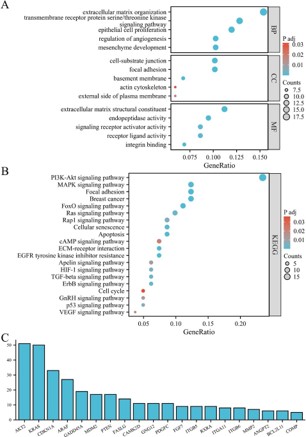
We performed gene functional enrichment analysis on the above 118 candidate genes. GO enrichment analysis (p<0.05) revealed a remarkable influence of 50 MF, including ECM structural constituent, endopeptidase activity, receptor ligand activity, signaling receptor activator activity, and integrin binding. Additionally, 34 CC such as focal adhesion, cell-substrate junction, basement membrane, external side of plasma membrane, and actin cytoskeleton had notable influences. Furthermore, significant effects were seen in 701 BP, including ECM organization, transmembrane receptor protein serine/threonine kinase signaling pathway, epithelial cell proliferation, mesenchyme development, and regulation of angiogenesis showed remarkable impacts (Figure 4A).
At the same time, the results of KEGG enrichment analysis (p<0.05) showed 118 candidate genes involved in 69 signaling pathways involving the aforementioned identified pathways related to chemoresistance and CAFs in breast cancer, such as FoxO signaling pathway, Rap1 signaling pathway, MAPK signaling pathway, ErbB signaling pathway, and PI3K-Akt signaling pathways (Figure 4B), and therefore, these pathways were considered as the crucial pathways in CAFs-mediated breast cancer drug resistance.
To further identify the crucial genes, we plot the top 20 genes based on their degree of enrichment in KEGG, including AKT2, KRAS, CDKN1A, ARAF, GADD45A, MDM 2, PTEN, FASLG, CAMK2D, GNG 12, PDGFC, FGF7, ITGB 5, RXRA, ITGA11, ITGB 6, MMP 2, ANGPT2, BCL2L11, and COMP (Figure 4C).
Construction of PPI network of candidate genes
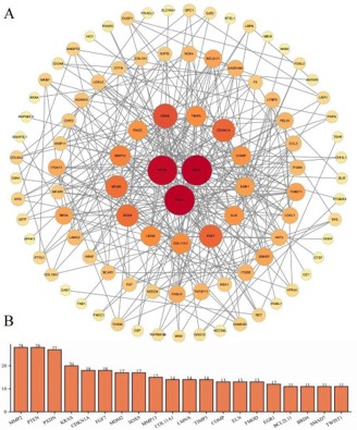
We restricted the species to human in the String database and imported these 118 candidate genes in order to investigate the PPI network of the proteins encoded by these genes in more detail. As can be seen in (Figure 5A), there were 329 edges and 118 nodes (PPI enrichment p value <1.0e-16) that demonstrated the correlations between proteins. The number of additional genes in the PPI network that interacted with the candidate gene was known as core degree value. Based on degree value, the top 20 genes were MMP2, PTEN, PXDN, KRAS, CDKN1A, FGF7, MDM2, SOX9, MMP13, COL11A1, LMNA, TIMP3, COMP, ELN, FMOD, EGR1, BCL2L11, BRD4, SMAD7, and TWIST1 (Figure 5B).
KRAS, COMP, PTEN, CDKN1A, and BCL2L11 are crucial genes regulating chemoresistance
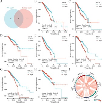
Eight crucial genes (MMP2, PTEN, KRAS, CDKN1A, FGF7, MDM2, COMP, and BCL2L11) were obtained from the intersection of PPI-top20 and KEGG-top20 based on the degree of enrichment and degree value (Figure 6A).
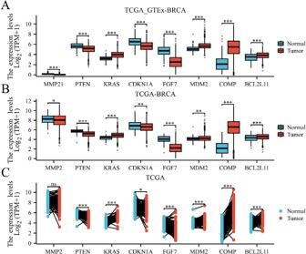
Meanwhile, we compared the mRNA expression of the above crucial genes in TCGA samples, TCGA_GTEx samples, and TCGA paired samples using BRCA expression profiling data provided by TCGA, and the results showed that KRAS, MDM2, COMP, and BCL2L11 were up-regulated, while MMP2, PTEN, CDKN1A, and FGF7 were down-regulated (Supplementary Figure 1A-C).
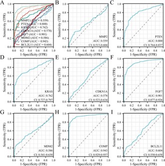
In addition, we plotted the receiver operating characteristic (ROC) curve of the above genes based on clinical information from the TCGA-BRCA dataset and the UCSC Xena database, and the results showed that the AUC of the above genes were all greater than 0.5. The AUC of the genes MMP2, CDKN1A, MDM2, and BCL2L11 were in the range of 0.5~0.7, with low diagnostic accuracy; the AUC of genes PTEN and KRAS were in the range of 0.7~0.9, with some diagnostic accuracy; and the AUC of genes FGF7 and COMP were >0.9, with high diagnostic accuracy (Supplementary Figure 2A-I).
The UCSC Xena database was used to analyze the relationship between the expression of the above 8 genes and the OS, DSS, and PFI of 1098 breast cancer patients in the TCGA-BRCA dataset. The results showed that KRAS, PTEN, CDKN1A were significantly correlated with OS, COMP, BCL2L11 were significantly correlated with DSS, and KRAS as well as COMP were significantly correlated with PFI (Figure 6B-H). Therefore, we identified KRAS, COMP, PTEN, CDKN1A, and BCL2L11 as crucial genes for chemoresistance in breast cancer.
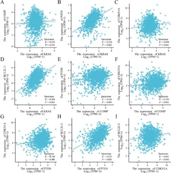
Finally, the correlation of the expression of the above genes was analyzed based on the TCGA-BRCA dataset, and we found that there was a positive correlation between the expression of the above crucial genes (Figure 6I), as detailed in Supplementary Figure 3A-I.
Crucial genes associated with clinical features
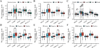
Based on the clinical information from TCGA-BRCA dataset and the UCSC Xena database, we further explored the correlation between the crucial genes and the clinical characteristics of breast cancer, including menstrual status, anatomical site, PAM50, and ER/PR/HER2 status (Figure 7A-I). The results demonstrated that in breast cancer patients, the expression of PTEN and BCL2L11 were linked to menstrual status. The expression of COMP was correlated with the anatomical site, with higher expression levels in right than left breast cancer. The expression of KRAS and COMP were correlated with different breast cancer subtypes. The expression of KRAS, COMP, PTEN, CDKNA1, and BCL2L11 were related to the status of ER and PR expression, while the expression of COMP was related to HER2 expression status.
Immune infiltration abundance of crucial genes
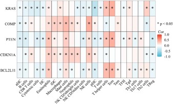
Given the interaction of CAFs with immune cells, we further investigated the correlation between crucial genes and the infiltration abundance of major immune cell types, including 24 immune cells like B cells, CD8+ T cells, macrophages, neutrophils, and dendritic cells. The results revealed that KRAS, COMP, PTEN, CDKN1A, and BCL2L11 were partially positively and partially negatively associated with immune cells. For instance, COMP and CDKN1A showed a positive correlation with CD8+ T cells infiltration but a negative correlation with B cells infiltration, while PTEN and BCL2L11 were positively associated with Natural Killer cells (NKs) infiltration but negatively associated with regulatory T cells (Tregs) infiltration (Figure 8).
Discussion
Taxanes is effective in treating breast cancer with few adverse reactions, and is often used as a first-line chemotherapeutic agent for adjuvant and neoadjuvant therapy in advanced metastatic breast cancer. However, the emergence of drug resistance is one of the important reasons for clinical treatment failure [19]. With a more in-depth understanding of the TME, it has also been demonstrated to be a crucial factor in the induction of chemotherapy resistance [20]. CAFs are the most numerous cellular components in the TME that can mediate cancer cell chemoresistance, thereby facilitating cancer development, invasion and metastasis [21,22]. For example, CAFs are reported as a key factor in chemoresistance in gastric and pancreatic cancers [23,24]. However, the particular mechanisms of CAFs’ function in chemoresistance in breast cancer remain unknown.
Studies have shown that CAFs are involved in inducing chemoresistance in breast cancer, and targeting CAFs may be a promising approach to overcome resistance [25,26]. For example, inhibition of interferon signaling prevents CAFs-dependent protection of breast cancer cells from chemotherapy [27]. Moreover, CAFs promotes resistance to cisplatin and DTX in locally advanced breast cancer patients and predict poor prognosis [28]. ATL-1 downregulates CTGF expression in fibroblasts and decreases the ability of breast cancer cells to convert fibroblasts into CAFs, thereby increasing the sensitivity of TNBC cells to PTX [29].
In our study, we performed bioinformatics analyses identifying crucial genes and pathways associated with CAFs in breast cancer chemoresistance to find molecular markers associated with breast cancer prognosis. As a result, this study revealed that crucial signaling pathways such as FoxO, Rap 1, MAPK, ErbB, PI3K-Akt and crucial genes such as KRAS, COMP, PTEN, CDKN1A, and BCL2L11s are important for the regulation of breast cancer chemoresistance, suggesting that these signaling pathways and genes are expected as potential intervention strategies for targeting CAFs to improve taxanes resistance, and provide more effective individualized treatment options for breast cancer patients.
FoxO (Forkhead box O) is an important transcription factor that is indispensable in the regulation of the cell cycle and apoptosis [30]. The FoxO signaling pathway is mainly regulated by PKB/Akt and MAPK [31]. Upon phosphorylation by Akt or other kinases, the ability of FoxO to bind DNA is inhibited, losing the ability to initiate transcription of target genes for apoptosis and cell cycle arrest [32]. In breast cancer, this pathway is frequently dysregulated, leading to sustained Akt activation and FoxO inactivation, which contributes to evasion of chemotherapy-induced apoptosis, leading to chemoresistance. For example, miR-222 mediates doxorubicin resistance in breast cancer cells by regulating the PTEN/Akt/FoxO1 signaling pathway [33]. In addition, FoxO3a inhibits the development of tamoxifen-resistant breast cancer by increasing the expression of integrin α5 [34] and plays an important role in regulating the drug sensitivity of breast cancer to epirubicin and PTX [35]. These findings are consistent with our findings.
ErbB belongs to the epidermal growth factor receptor family, and the human ErbB family consists of four members, ErbB1 (EGFR), ErbB2 (HER2), ErbB3, and ErbB4, which play key roles in regulating the processes of cell proliferation, migration, and differentiation [36]. ErbB exerts its effects by initiating downstream signaling cascades that affect various cellular processes, among which, the PI3K/Akt and MAPK signaling pathways are the most important [37]. In breast cancer, especially in HER2-positive and TNBC subtypes, over-activation of the ErbB pathway is often associated with resistance to conventional chemotherapeutic agents and poor prognosis. Generally, abnormally activated Akt in HER2positive breast cancer, which plays a major role in resistance to anti-HER 2 therapy via the PI3K/Akt pathway [38]. EGFR overexpression is also common in TNBC, leading to resistant by a similar mechanism [39].
Rap 1, also known as the Ras-associated protein 1. Rap 1 signaling activation plays a critical role in inducing breast cancer formation and progression to malignancy [40]. Upregulation of Rap 1 signaling leads to increased adhesion of tumor cells to the ECM, promoting the metastasis of breast cancer [41,42]. Furthermore, this process is essential for protecting tumor cells from chemotherapy-induced apoptosis, as increased adhesion can provide mechanical resistance to cell detachment during chemotherapy. For example, inhibition of the Rap 1 signaling in breast cancer can lead to attenuation of the malignant properties of breast cancer cells and chemotherapeutic resistance to doxorubicin [43]. Moreover, another interesting finding is that activating Rap 1 signaling enhances breast cancer migration and invasion but makes it more sensitive to rapamycin and PTX response [44].
The MAPK and PI3K-Akt signaling pathways have long been shown to be involved in breast cancer drug resistance in several studies. For example, activation of the MAPK signaling pathway in breast cancer has been experimentally shown to favor the development of resistance to cisplatin [45]. Modulation of the ERK/ MAPK signaling pathway promotes triple-negative breast cancer cell proliferation, invasion and DTX resistance [46,47], in addition to driving ECM degradation and PTX chemoresistance [48]. Whereas, activation of FAK/PI3K/Akt signaling pathway and IGF1RPI3K-Akt axis in breast cancer cells both induce cisplatin resistance and breast cancer progression [49,50]. Inhibition of tamoxifen and PTX therapeutic responsiveness by PI3K/Akt signaling in breast cancer has also been confirmed by studies [51,52].
Since most of the above pathways can function by regulating the MAPK and PI3K-Akt signaling pathways, we focused on the importance of these two in chemoresistance of CAFs in breast cancer. It was shown that IL 32 derived from CAFs enhances EMT and tumor cell invasion by activating the p38/MAPK signaling pathway in breast cancer [53]. Breast cancer cells can secrete TGFβ1, which in turn upregulates the p44/42 MAPK signaling pathway, leading to the chemoresistance of CAFs [54]. In addition, autocrine IGF2 from CAFs in breast cancer activates PI3K/Akt signaling by binding to IGF1 receptor on CAFs, promotes the interaction between CAFs and T cells, mediating immune escape, and ultimately leads to reduced efficacy of anti-programmed death 1 therapy [55]. CAFs can also induce trastuzumab resistance in HER2-positive breast cancer through amplification of cancer stem cells and activation of multiple pathways (NF-κ B, JAK/STAT3, and PI3K/Akt) [56].
However, the study of CAFs mediating PTX resistance in breast cancer through the above signaling pathways has not been reported, so further investigation of the potential relationship between CAFs and these related pathways may be an important direction to address PTX resistance clinically.
Moreover, we found that the crucial genes KRAS, COMP, PTEN, CDKN1A, BCL2L11, which are highly enriched in these signaling pathways and are closely related to the clinical prognosis of patients, are abnormal expression in breast cancer tissues, and correlate with CAFs and their mediated development of resistance to paclitaxel, which is a potential target for targeting CAFs to improve paclitaxel resistance.
KRAS is the most common RAS mutation and one of the most common oncogenic mutations. In breast cancer, KRAS mediate resistance to chemotherapeutic agents. For example, KRAS mutations in breast cancer reduce responsiveness to drug treatment with trastuzumab [57]. Furthermore, the emergence of KRAS mutations led to the generation of palbociclib resistance in metastatic breast cancer patients who received palbociclib combined with fulvestrant as a first-line treatment regimen [58]. While a cell experiment showed that the expression level of KRAS changed significantly in various cell lines before and after PTX treatment, suggesting its involvement in the drug response of PTX and may be apotential mechanism to overcome PTX resistance [59]. The KRAS protein was once considered a therapeutically refractory target due to its «undruggable», but in recent years, specific inhibitors against KRAS (G12C) mutations, such as Adagrasib and Sotorasib have been approved by the FDA and have shown promising efficacy in clinical trials [60,61]. They may be expected to be used in the treatment of breast cancer in the future.
COMP (Cartilage Oligomeric Matrix Protein) is an important non-collagenous ECM protein. It functions as a signaling regulator in maintaining the structural integrity of the ECM and is involved in various biological processes including cell adhesion, proliferation and differentiation [62]. The expression of COMP is usually higher in breast cancer tissues than in normal tissues and correlates with lymph node metastasis and poor prognosis [63], which is in line with our finding that high COMP expression is associated with poor TNM stage in breast cancer. Moreover, in metastatic patients treated with taxanes, the median survival of patients with high levels of serum COMP detected in the disease metastasis period was shorter [64], suggesting that COMP is associated with chemotherapy resistance involved such as increased cancer cell survival and apoptosis inhibition. It have also been found that COMP could inhibit the activity of calpain, thus reducing the activation of apoptosis-related proteins and enhance the viability of tumor cells by regulating the calcium homeostasis and apoptosis pathway, leading to resistance to a variety of chemotherapeutic drugs [65]. This is consistent with our findings.
PTEN is a key tumor suppressor gene that mainly dephosphorylates PIP3 through its lipid phosphatase activity, which in turn inhibits the activation of the PI3K/Akt signaling pathway [66]. It significantly affects the genomic stability, cell cycle of breast cancer, thereby promoting breast cancer development [67,68]. In addition, PTEN has been found to be involved in breast cancer drug resistance. For example, PTEN/Akt/mTOR signaling facilitates the promotion of a doxorubicin-resistant phenotype in breast cancer [69]. Deletion of PTEN may also create an immunosuppressive microenvironment by affecting the number of tumor-infiltrating lymphocytes, leading to immune escape and resistance to antiPD-1/PD-L1 therapy [70]. While the loss of function of PTEN has also been shown to be involved in regulating the PTX resistance of breast cancer by activating the PI3K/Akt pathway through multiple mechanisms [71,72]. These studies highlight the need for restoring PTEN function as a potential strategy to overcome drug resistance in breast cancer. For example, PTX combination therapy has shown promising in clinical trials for the treatment of advanced TNBC with altered PTEN [73]. CCDKN1A (Cyclin-Dependent Kinase Inhibitor 1A), also known as p21, is an important cell cycle regulator that plays a key role in the G1/S transition of the cell cycle, ensuring DNA integrity and normal cell division [74]. It has been found that p21 has a dual role in breast cancer as both a tumor suppressor and an oncogene, with specific mechanisms that are complex and dependent on the cellular environment and signaling pathways [75]. Abnormally expressed p21 in breast cancer protects cancer cells from the cytotoxic effects of chemotherapeutic drugs. For example, in the state of doxorubicin treatment, cell colony ability to form or undergo apoptosis is significantly affected by changes in p21 expression, and P21 expression protects MDA-MB-231 cells from inhibition of cell proliferation caused by doxorubicin [76]. Moreover, early studies have confirmed that p21 is important in mediating PTX resistance in breast cancer [77,78].
BCL2L11 (BCL-2 like 11), also known as BIM, is a pro-apoptotic protein and plays a key role in the mitochondria-dependent apoptotic pathway [79]. BIM has an important role in chemotherapy resistance in breast cancer, and its expression level and functional status directly affect the efficacy of chemotherapeutic agents. It was shown that the protein-protein interaction of Hsp 70 with BIM protects ERα-positive breast cancer from tamoxifen-induced apoptosis by binding and stabilizing ERα36 [80]. BIM is involved in regulating chemosensitivity of PRKCQ to PTX and doxorubicin treatments, and inhibition of BIM hinders chemotherapy-induced cell apoptosis [81]. Moreover, BIM regulates PTX-induced apoptosis through the mitochondrial pathway, significantly affecting the sensitivity of breast cancer cells to PTX, and is a potential target to overcome chemoresistance [82].
CAFs can interact with immune cells in the TME and significantly affect tumor progression and chemoresistance. CAFs can mediate immune escape from TNBC by secreting chemokines, recruiting monocytes and promoting their transformation into lipidassociated macrophages with immunosuppressive functions, and inhibiting T cell activation and proliferation [83]. It has also been shown that lymphocyte infiltration abundance is closely related to TNBC survival, which can be used as an early prognostic indicator and a predictor of TNBC response to chemotherapy [84].
Moreover, CAFs can inhibit the infiltration of CD8+ T cells and promote the resistance of tumor cells to immunotherapy by blocking the immune checkpoints [85]. Based on these findings, we hypothesize that crucial genes may regulate CAFs and influence the generation of chemoresistance in breast cancer through their interaction with immune cells, ultimately affecting patient survival. Our findings tentatively support this speculation. These findings not only reveal the central role of CAFs and immune cells in breast cancer chemoresistance, but also provide a theoretical basis for the development of therapeutic strategies targeting TME. In addition, some studies have found that the above genes and pathways could affect the chemotherapy efficacy of breast cancer through interaction. Two key signaling pathways for cell growth regulation, Ras/Raf/MEK/ERK and PI3K/PTEN/Akt/mTOR, can be activated by chemotherapeutic agents and are thus involved in the regulation of chemoresistance [86]. While sustained EGFR/ KRAS/SIAH pathway activation drives chemoresistance and early tumor recurrence in triple negative breast cancer [87].
However, studies involving the above genes and pathways in breast cancer chemotherapy resistance, especially involving CAFs, have not been reported and need further study.
In this study, we performed bioinformatics analysis to identify genes and pathways associated with CAFs and breast cancer chemoresistance based on public databases. We found that breast cancer resistance may be associated with CAFs, suggesting that CAF-related genes and pathways may serve as a potent tumor biomarker and possible therapeutic target, and providing direction for future mechanistic studies. Although the genes and pathways in the results have been extensively studied, most studies have focused on other biological roles, and they have not been reported in previous studies as chemotherapy resistance genes associated with CAFs. Therefore, they are still valuable to study as potential chemotherapeutic resistance genes associated with CAFs.
Although we merged information from several databases to make our findings accurate and reliable, some limitations of this study are inevitable. First of all, the limitation of the sample size in the relevant datasets leads to the underrepresentation of the sample, and more diverse samples and data are needed in the future to further support our conclusions. Therefore, the interaction between tumor cells and CAFs depends not only on shared genes but also on the correlation of gene expression between these two components, which we cannot currently demonstrate with publicly available data. Finally, the conclusions of this study are based on the results of bioinformatics analyses of public databases, and the real role of tumor-associated fibroblasts in chemotherapy resistance needs to be confirmed by more experiments. More studies of CAFs and chemotherapy resistance in breast cancer may contribute to giving a clear answer.
Therefore, targeted therapies targeting these genes may be very promising as an anti-chemotherapy resistance strategy for breast cancer patients. However, further research is necessary to confirm the findings prior to clinical translation, particularly regarding the potential of CAF-based targeted treatment to overcome breast cancer chemoresistance.
Conclusion
In conclusion, our study indicated that that FoxO, Rap1, MAPK, ErbB, PI3K-Akt signaling pathways may be the crucial pathways for CAFs in chemoresistance in breast cancer, while KRAS, COMP, PTEN, CDKN1A, and BCL2L11 may be crucial genes. This work offered a new perspective on the progression of treatment targets for breast cancer therapy based on CAFs as well as a novel molecular theory for the potential mechanisms by which CAFs mediate the effects of chemoresistance in breast cancer.
Declarations
Ethics approval and consent to participate: Ethics approval and patient informed consent were not needed since the study complied with TCGA and UCSC published guidelines.
Availability of data and materials: The datasets used and analyzed during the current study are available in the GEO database (https://www.ncbi.nlm.nih.gov/gds/), GeneCards database (https://www.genecards.org/), TCGA website (https://portal.gdc. cancer.gov/), UCSC Xena database (http://xena.ucsc.edu/), and String database (https://cn.string-db.org/).
Declaration of interests: The authors declare that they have no known competing financial interests or personal relationships that could have appeared to influence the work reported in this paper.
Author contributions: Li Wang: Data curation, Formal analysis, Investigation, Methodology, Visualization, Writing – original draft, Writing – review & editing. Han Xiao: Investigation, Methodology, Validation, Writing – original draft. Xiaoling Leng: Methodology, Project administration, Supervision, Writing – review & editing.
Guofu Huang: Conceptualization, Project administration, Supervision, Writing – review & editing.
Funding: This study was approved by the Xinjiang Uygur Autonomous Region Natural Science Fund [2022D01C301] and Cultivation Project of National Natural Science Foundation of China in The Tenth Affiliated Hospital, Southern Medical University (Dongguan People’s Hospital) [Z202410].
References
- Bray F, Laversanne M, Sung H, Ferlay J, Siegel RL, Soerjomataram I, et al. Global cancer statistics 2022: GLOBOCAN estimates of incidence and mortality worldwide for 36 cancers in 185 countries. CA Cancer J Clin. 2024; 74: 229–63.
- Dan VM, Raveendran RS, Baby S. Resistance to Intervention: Paclitaxel in Breast Cancer. Mini Rev Med Chem. 2021; 21: 1237–68.
- Hinshaw DC, Shevde LA. The Tumor Microenvironment Innately Modulates Cancer Progression. Cancer Res. 2019; 79: 4557–66.
- Terceiro LEL, Edechi CA, Ikeogu NM, Nickel BE, Hombach-Klonisch S, Sharif T, et al. The Breast Tumor Microenvironment: A Key Player in Metastatic Spread. Cancers. 2021; 13: 4798.
- Park D, Sahai E, Rullan A. SnapShot: Cancer-Associated Fibroblasts. Cell. 2020; 181: 486-486.e1.
- Qiao A, Gu F, Guo X, Zhang X, Fu L. Breast cancer-associated fibroblasts: their roles in tumor initiation, progression and clinical applications. Front Med. 2016; 10: 33–40.
- Houthuijzen JM, Jonkers J. Cancer-associated fibroblasts as key regulators of the breast cancer tumor microenvironment. Cancer Metastasis Rev. 2018; 37: 577–97.
- Fiori ME, Di Franco S, Villanova L, Bianca P, Stassi G, De Maria R. Cancer-associated fibroblasts as abettors of tumor progression at the crossroads of EMT and therapy resistance. Mol Cancer. 2019; 18: 70.
- Avalle L, Raggi L, Monteleone E, Savino A, Viavattene D, Statello L, et al. STAT3 induces breast cancer growth via ANGPTL4, MMP13 and STC1 secretion by cancer associated fibroblasts. Oncogene. 2022; 41: 1456–67.
- Huang M, Fu M, Wang J, Xia C, Zhang H, Xiong Y, et al. TGF-β1activated cancer-associated fibroblasts promote breast cancer invasion, metastasis and epithelial-mesenchymal transition by autophagy or overexpression of FAP-α. Biochem Pharmacol. 2021; 188: 114527.
- Hu G, Xu F, Zhong K, Wang S, Huang L, Chen W. Activated Tumorinfiltrating Fibroblasts Predict Worse Prognosis in Breast Cancer Patients. J Cancer. 2018; 9: 3736–42.
- Plava J, Cihova M, Burikova M, Matuskova M, Kucerova L, Miklikova S. Recent advances in understanding tumor stroma-mediated chemoresistance in breast cancer. Mol Cancer. 2019; 18: 67.
- Gao Y, Li X, Zeng C, Liu C, Hao Q, Li W, et al. CD63 + Cancer-Associated Fibroblasts Confer Tamoxifen Resistance to Breast Cancer Cells through Exosomal miR-22. Advanced Science. 2020; 7: 2002518.
- Fang L, Liu Q, Cui H, Zheng Y, Wu C. Bioinformatics Analysis Highlight Differentially Expressed CCNB1 and PLK1 Genes as Potential Anti-Breast Cancer Drug Targets and Prognostic Markers. Genes. 2022; 13: 654.
- An G, Feng L, Hou L, Li X, Bai J, He L, et al. A bioinformatics analysis of zinc finger protein family reveals potential oncogenic biomarkers in breast cancer. Gene. 2022; 828: 146471.
- Dai J, Zhu B, Lin W, Gao H, Dai H, Zheng L, et al. Identification of prognostic significance of BIRC5 in breast cancer using integrative bioinformatics analysis. Bioscience Reports. 2020; 40: BSR20193678.
- Cancer Genome Atlas Research Network, Weinstein JN, Collisson EA, Mills GB, Shaw KRM, Ozenberger BA, et al. The Cancer Genome Atlas Pan-Cancer analysis project. Nat Genet. 2013; 45: 1113–20.
- T B, Se W, P L, C E, If K, M T, et al. NCBI GEO: archive for functional genomics data sets--update. Nucleic Acids Research. 2013; 41.
- Abu Samaan TM, Samec M, Liskova A, Kubatka P, Büsselberg D. Paclitaxel’s Mechanistic and Clinical Effects on Breast Cancer. Biomolecules. 2019; 9: 789.
- Mehraj U, Dar AH, Wani NA, Mir MA. Tumor microenvironment promotes breast cancer chemoresistance. Cancer Chemother Pharmacol. 2021; 87: 147–58.
- Jena BC, Das CK, Bharadwaj D, Mandal M. Cancer associated fibroblast mediated chemoresistance: A paradigm shift in understanding the mechanism of tumor progression. Biochim Biophys Acta Rev Cancer. 2020; 1874: 188416.
- Bu L, Baba H, Yasuda T, Uchihara T, Ishimoto T. Functional diversity of cancer-associated fibroblasts in modulating drug resistance. Cancer Sci. 2020; 111: 3468–77.
- Hu C, Xia R, Zhang X, Li T, Ye Y, Li G, et al. circFARP1 enables cancerassociated fibroblasts to promote gemcitabine resistance in pancreatic cancer via the LIF/STAT3 axis. Mol Cancer. 2022; 21: 24.
- Zhang H, Deng T, Liu R, Ning T, Yang H, Liu D, et al. CAF secreted miR-522 suppresses ferroptosis and promotes acquired chemo-resistance in gastric cancer. Mol Cancer. 2020; 19: 43.
- Zeng W, Xiong L, Wu W, Li S, Liu J, Yang L, et al. CCL18 signaling from tumor-associated macrophages activates fibroblasts to adopt a chemoresistance-inducing phenotype. Oncogene. 2023; 42: 224–37.
- Fan G, Yu B, Tang L, Zhu R, Chen J, Zhu Y, et al. TSPAN8+ myofibroblastic cancer-associated fibroblasts promote chemoresistance in patients with breast cancer. Sci Transl Med. 2024; 16: eadj5705.
- Broad RV, Jones SJ, Teske MC, Wastall LM, Hanby AM, Thorne JL, et al. Inhibition of interferon-signalling halts cancer-associated fibroblast-dependent protection of breast cancer cells from chemotherapy. Br J Cancer. 2021; 124: 1110–20.
- Al-Tweigeri T, AlRaouji NN, Tulbah A, Arafah M, Aboussekhra M, AlMohanna F, et al. High AUF1 level in stromal fibroblasts promotes carcinogenesis and chemoresistance and predicts unfavorable prognosis among locally advanced breast cancer patients. Breast Cancer Res. 2022; 24: 46.
- Wang M, Li X-Z, Zhang M-X, Ye Q-Y, Chen Y-X, Chang X. Atractylenolide-I Sensitizes Triple-Negative Breast Cancer Cells to Paclitaxel by Blocking CTGF Expression and Fibroblast Activation. Front Oncol. 2021; 11: 738534.
- Bach D-H, Long NP, Luu T-T-T, Anh NH, Kwon SW, Lee SK. The Dominant Role of Forkhead Box Proteins in Cancer. Int J Mol Sci. 2018; 19: 3279.
- Roy SK, Srivastava RK, Shankar S. Inhibition of PI3K/AKT and MAPK/ ERK pathways causes activation of FOXO transcription factor, leading to cell cycle arrest and apoptosis in pancreatic cancer. J Mol Signal. 2010; 5: 10.
- X Z, N T, Tj H, Ak R. Akt, FoxO and regulation of apoptosis. Biochimica et Biophysica Acta. 2011: 1813.
- Shen H, Wang D, Li L, Yang S, Chen X, Zhou S, et al. MiR-222 promotes drug-resistance of breast cancer cells to adriamycin via modulation of PTEN/Akt/FOXO1 pathway. Gene. 2017; 596: 110–8.
- Ricci E, Fava M, Rizza P, Pellegrino M, Bonofiglio D, Casaburi I, et al. FoxO3a Inhibits Tamoxifen-Resistant Breast Cancer Progression by Inducing Integrin α5 Expression. Cancers. 2022; 14: 214.
- Khongkow M, Olmos Y, Gong C, Gomes AR, Monteiro LJ, Yagüe E, et al. SIRT6 modulates paclitaxel and epirubicin resistance and survival in breast cancer. Carcinogenesis. 2013; 34: 1476–86.
- Yarden Y, Sliwkowski MX. Untangling the ErbB signalling network. Nat Rev Mol Cell Biol. 2001; 2: 127–37.
- Ghayad SE, Vendrell JA, Ben Larbi S, Dumontet C, Bieche I, Cohen PA. Endocrine resistance associated with activated ErbB system in breast cancer cells is reversed by inhibiting MAPK or PI3K/Akt signaling pathways. Int J Cancer. 2010; 126: 545–62.
- L P, J L, Q X, Z G, M Y, X W, et al. HER2/PI3K/AKT pathway in HER2-positive breast cancer: A review. Medicine. 2024: 103.
- Hsu JL, Hung MC. The role of HER2, EGFR, and other receptor tyrosine kinases in breast cancer. Cancer Metastasis Rev. 2016; 35: 575–88.
- Itoh M, Nelson CM, Myers CA, Bissell MJ. Rap1 integrates tissue polarity, lumen formation, and tumorigenic potential in human breast epithelial cells. Cancer Res. 2007; 67: 4759–66.
- McSherry EA, Brennan K, Hudson L, Hill ADK, Hopkins AM. Breast cancer cell migration is regulated through junctional adhesion molecule-A-mediated activation of Rap1 GTPase. Breast Cancer Res. 2011; 13: R31.
- Zhang Y-L, Wang R-C, Cheng K, Ring BZ, Su L. Roles of Rap1 signaling in tumor cell migration and invasion. Cancer Biol Med. 2017; 14: 90–9.
- S T, Z B, Y L, Y G, J Z, Y Z, et al. Exosomes Derived from Tumor Cells Initiate Breast Cancer Cell Metastasis and Chemoresistance through a MALAT1-Dependent Mechanism. Journal of Oncology. 2022: 2022.
- Zhang J, Guo F, Li C, Wang Y, Wang J, Sun F, et al. Loss of TTC17 promotes breast cancer metastasis through RAP1/CDC42 signaling and sensitizes it to rapamycin and paclitaxel. Cell Biosci. 2023; 13: 50.
- Feng C, Li P, Liu P, Wang B, Li J, Liu P. LMAN2 Promotes Breast Cancer Tumorigenesis and Drug Resistance by Interacting with MAPK9 via Activation of the MAPK Pathway. Cancer Med. 2024; 13: e70448.
- Wang B, Xing A-Y, Li G-X, Liu L, Xing C. SNHG14 promotes triplenegative breast cancer cell proliferation, invasion, and chemoresistance by regulating the ERK/MAPK signaling pathway. IUBMB Life. 2024; 76: 1295–308.
- Zhao P, Ma W, Hu Z, Zang L, Tian Z, Zhang K. Filamin A (FLNA) modulates chemosensitivity to docetaxel in triple-negative breast cancer through the MAPK/ERK pathway. Tumour Biol. 2016; 37: 5107–15.
- Wu J, Li J, Xu H, Qiu N, Huang X, Li H. Periostin drives extracellular matrix degradation, stemness, and chemoresistance by activating the MAPK/ERK signaling pathway in triple-negative breast cancer cells. Lipids Health Dis. 2023; 22: 153.
- Luo J, Yao J-F, Deng X-F, Zheng X-D, Jia M, Wang Y-Q, et al. 14, 15EET induces breast cancer cell EMT and cisplatin resistance by upregulating integrin αvβ3 and activating FAK/PI3K/AKT signaling. J Exp Clin Cancer Res. 2018; 37: 23.
- Zhou L, Li H, Sun T, Wen X, Niu C, Li M, et al. HULC targets the IGF1R–PI3K-AKT axis in trans to promote breast cancer metastasis and cisplatin resistance. Cancer Letters. 2022; 548: 215861.
- Tian Y, Chen Z-H, Wu P, Zhang D, Ma Y, Liu X-F, et al. MIR497HGDerived miR-195 and miR-497 Mediate Tamoxifen Resistance via PI3K/AKT Signaling in Breast Cancer. Adv Sci (Weinh). 2023; 10: e2204819.
- Cai J, Chen S, Zhang W, Zheng X, Hu S, Pang C, et al. Salvianolic acid A reverses paclitaxel resistance in human breast cancer MCF7 cells via targeting the expression of transgelin 2 and attenuating PI3 K/Akt pathway. Phytomedicine. 2014; 21: 1725–32.
- Wen S, Hou Y, Fu L, Xi L, Yang D, Zhao M, et al. Cancer-associated fibroblast (CAF)-derived IL32 promotes breast cancer cell invasion and metastasis via integrin β3-p38 MAPK signalling. Cancer Lett. 2019; 442: 320–32.
- Chandra Jena B, Kanta Das C, Banerjee I, Das S, Bharadwaj D, Majumder R, et al. Paracrine TGF-β1 from breast cancer contributes to chemoresistance in cancer associated fibroblasts via upregulation of the p44/42 MAPK signaling pathway. Biochem Pharmacol. 2021; 186: 114474.
- Song D, Wu Y, Li J, Liu J, Yi Z, Wang X, et al. Insulin-like growth factor 2 drives fibroblast-mediated tumor immunoevasion and confers resistance to immunotherapy. J Clin Invest. 2024; 134: e183366.
- Mao Y, Zhang Y, Qu Q, Zhao M, Lou Y, Liu J, et al. Cancer-associated fibroblasts induce trastuzumab resistance in HER2 positive breast cancer cells. Mol Biosyst. 2015; 11: 1029–40.
- Patra S, Young V, Llewellyn L, Senapati JN, Mathew J. BRAF, KRAS and PIK3CA Mutation and Sensitivity to Trastuzumab in Breast Cancer Cell Line Model. Asian Pac J Cancer Prev. 2017; 18: 2209– 13.
- Raimondi L, Raimondi FM, Pietranera M, Di Rocco A, Di Benedetto L, Miele E, et al. Assessment of Resistance Mechanisms and Clinical Implications in Patients with KRAS Mutated-Metastatic Breast Cancer and Resistance to CDK4/6 Inhibitors. Cancers (Basel). 2021; 13: 1928.
- Asghari F, Haghnavaz N, Shanehbandi D, Khaze V, Baradaran B, Kazemi T. Differential altered expression of let-7a and miR-205 tumor-suppressor miRNAs in different subtypes of breast cancer under treatment with Taxol. Adv Clin Exp Med. 2018; 27: 941–5.
- Zhang Y, Li C, Xia C, Wah To KK, Guo Z, Ren C, et al. Adagrasib, a KRAS G12C inhibitor, reverses the multidrug resistance mediated by ABCB1 in vitro and in vivo. Cell Commun Signal. 2022; 20: 142.
- Vishakha S, Navneesh N, Kurmi BD, Gupta GD, Verma SK, Jain A, et al. An Expedition on Synthetic Methodology of FDA-approved Anticancer Drugs (2018-2021). Anticancer Agents Med Chem. 2024; 24: 590–626.
- Cui J, Zhang J. Cartilage Oligomeric Matrix Protein, Diseases, and Therapeutic Opportunities. Int J Mol Sci. 2022; 23: 9253.
- Papadakos KS, Hagerling C, Rydén L, Larsson A-M, Blom AM. High Levels of Expression of Cartilage Oligomeric Matrix Protein in Lymph Node Metastases in Breast Cancer Are Associated with Reduced Survival. Cancers (Basel). 2021; 13: 5876.
- Papadakos KS, Darlix A, Jacot W, Blom AM. High Levels of Cartilage Oligomeric Matrix Protein in the Serum of Breast Cancer Patients Can Serve as an Independent Prognostic Marker. Front Oncol. 2019; 9: 1141.
- Hanitrarimalala V, Bednarska I, Murakami T, Papadakos KS, Blom AM. Intracellular cartilage oligomeric matrix protein augments breast cancer resistance to chemotherapy. Cell Death Dis. 2024; 15: 480.
- Papa A, Pandolfi PP. The PTENˉPI3K Axis in Cancer. Biomolecules. 2019; 9: 153.
- Bassi C, Fortin J, Snow BE, Wakeham A, Ho J, Haight J, et al. The PTEN and ATM axis controls the G1/S cell cycle checkpoint and tumorigenesis in HER2-positive breast cancer. Cell Death Differ. 2021; 28: 3036–51.
- Chen J, Sun J, Wang Q, Du Y, Cheng J, Yi J, et al. Systemic Deficiency of PTEN Accelerates Breast Cancer Growth and Metastasis. Front Oncol. 2022; 12: 825484.
- Yin Y, Wang X, Li T, Ren Q, Li L, Sun X, et al. MicroRNA-221 promotes breast cancer resistance to adriamycin via modulation of PTEN/Akt/mTOR signaling. Cancer Med. 2020; 9: 1544–52.
- Cretella D, Digiacomo G, Giovannetti E, Cavazzoni A. PTEN Alterations as a Potential Mechanism for Tumor Cell Escape from PD-1/ PD-L1 Inhibition. Cancers (Basel). 2019; 11: 1318.
- Geng W, Song H, Zhao Q, Dong K, Pu Q, Gao H, et al. miR-520h Stimulates Drug Resistance to Paclitaxel by Targeting the OTUD3PTEN Axis in Breast Cancer. Biomed Res Int. 2020; 2020: 9512793.
- Liu L, Meng T, Zheng X, Liu Y, Hao R, Yan Y, et al. Transgelin 2 Promotes Paclitaxel Resistance, Migration, and Invasion of Breast Cancer by Directly Interacting with PTEN and Activating PI3K/Akt/ GSK-3β Pathway. Mol Cancer Ther. 2019; 18: 2457–68.
- Pant S, Hamilton E, Ulahannan SV, Strauss JF, Braiteh FS, Huang M, et al. Phase 1b study of pan-AKT inhibitor vevorisertib alone or with paclitaxel or fulvestrant in PIK3CA/AKT/PTEN-mutated advanced solid tumors. Cancer. 2023; 129: 1919–29.
- Manousakis E, Miralles CM, Esquerda MG, Wright RHG. CDKN1A/ p21 in Breast Cancer: Part of the Problem, or Part of the Solution? Int J Mol Sci. 2023; 24: 17488.
- Kreis N-N, Louwen F, Yuan J. The Multifaceted p21 (Cip1/Waf1/ CDKN1A) in Cell Differentiation, Migration and Cancer Therapy. Cancers (Basel). 2019; 11: 1220.
- Huang P, Ouyang D-J, Chang S, Li M-Y, Li L, Li Q-Y, et al. Chemotherapy-driven increases in the CDKN1A/PTN/PTPRZ1 axis promote chemoresistance by activating the NF-κB pathway in breast cancer cells. Cell Commun Signal. 2018; 16: 92.
- Lv K, Liu L, Wang L, Yu J, Liu X, Cheng Y, et al. Lin28 mediates paclitaxel resistance by modulating p21, Rb and Let-7a miRNA in breast cancer cells. PLoS One. 2012; 7: e40008.
- Choi YH, Yoo YH. Taxol-induced growth arrest and apoptosis is associated with the upregulation of the Cdk inhibitor, p21WAF1/ CIP1, in human breast cancer cells. Oncol Rep. 2012; 28: 2163–9.
- O’Connor L, Strasser A, O’Reilly LA, Hausmann G, Adams JM, Cory S, et al. Bim: a novel member of the Bcl-2 family that promotes apoptosis. EMBO J. 1998; 17: 384–95.
- Song T, Zhang H, Zhao Q, Hu Z, Wang Z, Song Y, et al. Small molecule inhibitor targeting the Hsp70-Bim protein-protein interaction in estrogen receptor-positive breast cancer overcomes tamoxifen resistance. Breast Cancer Res. 2024; 26: 33.
- Byerly JH, Port ER, Irie HY. PRKCQ inhibition enhances chemosensitivity of triple-negative breast cancer by regulating Bim. Breast Cancer Res. 2020; 22: 72.
- Sun W-L, He L-Y, Liang L, Liu S-Y, Luo J, Lv M-L, et al. Ambra1 regulates apoptosis and chemosensitivity in breast cancer cells through the Akt-FoxO1-Bim pathway. Apoptosis. 2022; 27: 329–41.
- Timperi E, Gueguen P, Molgora M, Magagna I, Kieffer Y, Lopez-Lastra S, et al. Lipid-Associated Macrophages Are Induced by CancerAssociated Fibroblasts and Mediate Immune Suppression in Breast Cancer. Cancer Res. 2022; 82: 3291–306.
- Denkert C, von Minckwitz G, Darb-Esfahani S, Lederer B, Heppner BI, Weber KE, et al. Tumour-infiltrating lymphocytes and prognosis in different subtypes of breast cancer: a pooled analysis of 3771 patients treated with neoadjuvant therapy. Lancet Oncol. 2018; 19: 40–50.
- Jenkins L, Jungwirth U, Avgustinova A, Iravani M, Mills A, Haider S, et al. Cancer-Associated Fibroblasts Suppress CD8+ T-cell Infiltration and Confer Resistance to Immune-Checkpoint Blockade. Cancer Research. 2022; 82: 2904–17.
- Ja M, Ls S, Wh C, Sl A, Ra F, G M, et al. Ras/Raf/MEK/ERK and PI3K/ PTEN/Akt/mTOR cascade inhibitors: how mutations can result in therapy resistance and how to overcome resistance. Oncotarget. 2012; 3.
- Tang AH, Hoefer RA, Guye ML, Bear HD. Persistent EGFR/K-RAS/ SIAH pathway activation drives chemo-resistance and early tumor relapse in triple-negative breast cancer. Cancer Drug Resist. 2022; 5: 691–702.