Introduction
With the aging of the global population, there are more and more elderly breast cancer. As of 2016, more than 50% of breast cancer patients are over 60 years old [1]. Surgery combined with adjuvant chemotherapy and radiotherapy can effectively improve the survival rate of patients with breast cancer [2], but elderly breast cancer patients have their own characteristics, such as older age, decline of important organ function, more systemic accompanying diseases, poor surgical tolerance, etc., so there is no unified treatment plan for elderly breast cancer [3]. In addition, the damage of heart function caused by anthracycline toxicity is paid more attention by medical workers. Therefore, it is very important to find some accurate predictors for elderly breast cancer to identify their better prognosis, choose individualized treatment, and reduce the decline of quality of life caused by over treatment.
In recent years, many researchers have noticed the relationship between inflammation and tumorigenesis and development. Lymphocytes and Neutrophils are the most important effector cells, which play an important role in the body’s inflammatory response. Cytokines in Platelets, such as CD40L and interleukin (IL-1, IL-3, IL-6), also play an important role in inflammation and immune response. Many reports have shown that inflammatory indicators such as neutrophil/lymphocyte ratio (NLR), Platelet/ Lymphocyte Ratio (PLR), and Systemic Immune inflammatory Index (SII) are prognostic factors for for Lung cancer, Gastric cancer, Cholangiocarcinoma and other malignant tumors [4-6]. With the increase in age, the body’s chronic inflammatory response increases, and the inflammatory markers also increase accordingly [7], but whether inflammatory markers can play the same role in the prognosis evaluation of elderly cancer patients is still unclear. Therefore, the purpose of this study was to explore the prognostic significance of peripheral blood inflammatory markers in elderly patients with breast cancer, and to provide a reference value for oncologists in the treatment choice.
Materials and methods
Patient selection
Methods: A total of 260 elderly patients with breast cancer diagnosed at Guangxi Medical University Cancer Hospital between January 2003 and May 2018 were retrospectively analyzed. All patients signed informed consent for treatment and follow-up. All procedures conform to the ethical standards of the agency and the National Research Council, as well as the 1964 Helsinki declaration. This study was reviewed and approved by the ethics committee of Guangxi Medical University Cancer Hospital (Approval number: LW2021025)
Inclusion criteria: (A) All patients were diagnosed at least 65 years old; (B) All patients underwent radical surgery for breast cancer; (C) All patients were confirmed to have invasive ductal carcinoma by preoperative puncture and postoperative pathological diagnosis; (D) All patients had complete clinical records and follow-up data; (E) All peripheral blood samples were collected one week before treatment, and the inflammatory marker data were complete.
Exclusion criteria: (A) Receiving chemotherapy, radiotherapy, endocrine therapy, and targeted therapy before collecting peripheral blood samples; (B) Male breast cancer patients; (C) Patients with acute and chronic inflammatory diseases or autoimmune diseases such as pneumonia, systemic lupus erythematosus, and hepatitis; and (D) Patients with undetermined clinical data such as pathological stage and molecular typing; (E) Patients with distant metastasis defined before operation.
Classification criteria
TNM stages was based on the eighth edition of the American Joint Committee on Cancer (AJCC) and the Union for International Cancer Control (UICC) [8,9]. All patients in this study were stage IIII patients. The diagnosis of invasive ductal carcinoma in this study was based on the 2012 World Health Organization (WHO) breast tumor classification into grade I, II and III [10]. Molecular typing was based on St. Gallen international breast cancer treatment expert consensus in 2019 [11], which defines ER and PR expression >1% as positive, Ki-67 expression >14% as high expression, and it were divided into Luminal A, Luminal B1 (luminal B-like (HER2 negative)), Luminal B2 (luminal B-like (HER2 positive)), HER-2 overexpression type and Triple Negative Breast Cancer (TNBC).
Data collection
The clinical characteristics of the patients were collected, including age at first diagnosis, pathological grade, vascular cancer thrombus, clinical stage, molecular typing, surgical methods, endocrine therapy, and peripheral blood parameters. Peripheral blood was collected before neoadjuvant chemotherapy and surgery for the first time. Blood samples were collected with sterile EDTA tube and analyzed by bc-6900 automatic hematology analyzer. The main indexes analyzed were neutrophil (n), lymphocyte (L), platelet (P), and the corresponding inflammatory indexes NLR, PLR, SII. SII was defined as neutrophil count * platelet count / lymphocyte count.
Follow up
All patients who underwent surgery were defined as the starting point for calculating the survival time. They were reexamined every 3 months within 2 years, half a year after 2–5 years, and every 1 year after 5 years. During the study period, outpatient or telephone follow-up was conducted every half a year. The followup evaluation items mainly included symptom inquiry, physical examination, laboratory examination (blood routine, blood biochemistry, tumor markers, etc.), breast, liver, thyroid, gynecological ultrasound, and CT examination.
Statistical analysis
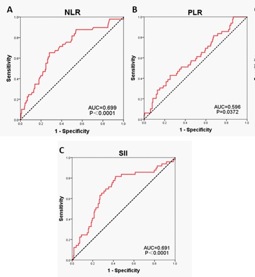
IBM SPSS software (version 24.0) and Empower stats software (version 2.2) were used to perform the data analysis. The optimal cut-off values of NLR, PLR and SII were 2.07, 185.27 and 428.44, respectively. Continuous variables were described by (mean±standard deviation) and analyzed by t test; categorical variables were described by [n (%); C2 test was used to evaluate the correlation between NLR, PLR, SII and clinical features. Kaplan Meier method and log rank test were used for single factor analysis survival analysis. The Overall Survival (OS) and Disease-Free Survival (DFS) of different variables were compared, DFS refers to the time from the operation time to the first local recurrence, distant metastasis, contralateral breast cancer, other secondary primary cancer or death from any cause. OS refers to the time from the operation time to death from any cause. Cox proportional hazard regression model was used in multivariate survival analysis. Variables with P < 0.05 in single factor analysis were included in Cox proportional hazard regression model to evaluate the independent correlation between risk ratio (HR) and 95% Confidence Interval (CI) of each factor DFS and OS of P<0.05 was set as statistically significant.
Results
A total of 260 elderly patients with breast cancer were included in this study. 227 of them had complete DFS and OS follow-up data. The subjects were followed up to July 2020, with an average follow-up time of 62 months (5~120 months). Among the 260 patients, the median age of initial diagnosis was 69 years old (65~92 years old), and 141 patients were less than 70 years old. There were 69 patients aged 70~74, 35 patients aged 75~79, and 15 patients aged 80 and above. The pathological types of 260 patients were invasive ductal carcinoma, of which 133 patients had histological grading data, including 8 cases (6.02%) in grade I, 69 cases (51.9%) in grade II and 56 cases (42.1%) in grade III; 155 patients had tumor vascular tumor thrombus data, including 112 cases (72.3%) with vascular tumor thrombus and 43 cases (27.7%) without vascular tumor thrombus. In clinical stage, 165 cases (63.46%) were stage II breast cancer, while 36 cases (13.85%) were stage I and 59 cases (22.69%) were stage III. There were 141 cases (54.23%) without axillary lymph node metastasis, 81 cases (31.15%) with 1~3 lymph nodes metastasis, 24 cases (9.23%) with 4~9 lymph nodes metastasis, and 14 cases (5.38%) with more than 9 lymph nodes metastasis. According to the sentinel lymph node status, 171 patients (65.77%) underwent modified radical mastectomy, 66(25.38%) underwent simple mastectomy plus sentinel lymph node biopsy, 21(8.08%) underwent breast conserving surgery, and 2(0.77%) underwent breast reconstruction. The most common molecular typing was luminal B1, with 94 cases (36.13%), followed by triple negative type, with 46 cases (17.69%). Among them, 168 cases were hormone receptor positive, 122 cases (72%) received endocrine therapy, and 46 cases (72%) did not receive endocrine therapy. Among the 122 patients who received endocrine therapy, more than 85% chose aromatase (AI) therapy.
Obtaining the optimal cut-off values of NLR, PLR and SII
NLR, PLR and SII were used as test variables to make ROC curve. The AUC of NLR, PLR and SII were 0.699 (P<0.001), 0.596 (P=0.037) and 0.691 (P<0.001), respectively. According to the highest Youden index, the cut-off points of NLR, PLR and SII are 2.04, 185.27 and 429.06. (Table 1, Figure). According to the cutoff value, NLR, PLR and SII were divided into NLR low expression group (169 cases), NLR high expression group (91 cases), PLR low expression group (186 cases), PLR high expression group (74 cases), SII low expression group (133 cases), SII high expression group (127 cases) (Table 2).
Correlation between clinical parameters and NLR, PLR, SII
In our study, the expression of NLR, PLR and SII had no significant difference in the overall population distribution (P>0.05) (Table 2).
Survival analysis
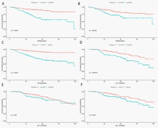
Single factor analysis of influencing factors of DFS and OS: In single factor analysis, age (P=0.018), histological grade (P=0.037), clinical stage (P<0.001), molecular typing (P= 0.006), NLR (P<0.001), PLR (P=0.006) and SII (P<0.001) were the influencing factors of DFS in 227 elderly patients with breast cancer (<0.05). Age (P=0.013), clinical stage (P<0.017), molecular typing (P<0.001), endocrine therapy (P=0.024), NLR (P<0.001) and SII (P=0.047) were the influencing factors of OS (<0.05). The low expression of NLR and SII was significantly correlated with better OS (Figure 2 & Table 3).
Cox proportional hazards regression model was used to analyze the independent prognostic factors of DFS and OS: In our study, Age, clinical stage, molecular subtype, NLR, PLR and SII were included in the Cox proportional hazards regression model of DFS. The results showed that clinical stage (HR=5.122, 95% CI 1.596-16.440, P=0.006), NLR (HR=2.340, 95% CI 1.089-5.028, P=0.029) and SII (HR=3.132, 95% CI 1.263~7.766, P=0.014) were an independent prognostic factor for DFS in elderly breast cancer. The risk of disease progression in clinical stage III patients was 5.122 times higher than that in stage I patients, the risk of disease progression in NLR high expression group was 2.34 times higher than that in NLR low expression group, and the risk of disease progression in SII high expression group was 3.132 times higher than that in low expression group. Our results showed that PLR could not independently predict DFS in elderly patients with breast cancer (HR=1.108, 95% CI 0.593-2.069, P=0.748) (Table 4).
Age, clinical stage, molecular classification, endocrine therapy drugs, NLR and SII were included in the Cox proportional hazards regression model. The results showed that age (HR=4.893, 95% CI 1.911-12.532, P=0.001), clinical stage (HR=3.215, 95% CI 1.030-10.034, P=0.017), HER2 overexpression (HR=4.017, 95% CI 1.911-12.532, P=0.001), TNBC (HR=5. 178, 95% CI 1. 850 ~ 14. 532, P=0.002), NLR (HR=2. 408, 95% CI 1. 417 ~ 4. 092, P=0.002) were significantly correlated with OS, The risk of death in patients over 80 years old was 4.893 times that in 65-69 years old, and the risk of death in patients with clinical stage III was 3.215 times that in patients with clinical stage I; the risk of death in patients with HER2 overexpression and TNBC was 4.017 times and 5.178 times that in patients with Luminal A, respectively; the risk of death in patients with high NLR expression was 2.408 times that in patients with low NLR expression. In addition, this study found that the impact of SII on OS in elderly breast cancer has a strong tendency (HR=1.711, 95% CI 1.000 ~ 2.928, P=0.05) (Table 4).
In subgroup analysis of molecular typing, high expression of NLR was an independent factor of poor DFs in luminal B1 patients (HR=6.148, 95% CI 1.255-30.111, P=0.025), while high expression of SII had a certain tendency in high recurrence risk (HR=4.983, 95% CI 0.788-31.513, P=0.088). High expression of NLR (P<0.001) and SII (P<0.001) were independent factors of poor OS in luminal B2 patients. High SII expression in TNBC patients was an independent factor for poor DFS (HR=12.143, 95% CI 1.151-128.090, P=0.038), but not for OS (HR=2.377, 95% CI 0.386-14.645, P=0.351).
Table 1: Bar chart of mean COX-2 expression for each treatment group with error bars as standard deviation.
| Variable |
AUC |
95%CI |
Z |
P |
Yoden index |
Cut-off point |
Sensitivity |
Specificity |
| NLR |
0.699 |
0.635~0.758 |
4.709 |
0.001* |
0.3722 |
2.04 |
65.31 |
71.91 |
| PLR |
0.596 |
0.529~0.660 |
2.084 |
0.037* |
0.1758 |
185.27 |
42.86 |
74.72 |
| SII |
0.691 |
0.625~0.751 |
4.343 |
0.001* |
0.3950 |
429.06 |
81.63 |
57.87 |
Remarks: *P<0.05
Table 2: Comparison of clinical characteristics of subjects with different inflammatory markers expression.
| Variables |
Total Patients (N=260) |
NLR |
PLR |
SII |
| Low (N=169) |
High (N=91) |
P value |
Low (N=186) |
High N=74) |
P value |
Low (N=133) |
High (N=127) |
P value |
| Age (years) |
260 |
|
|
0.094 |
|
|
0.377 |
|
|
0.643 |
| 65~69 |
141 |
98 |
43 |
100 |
41 |
71 |
70 |
| 70~74 |
69 |
46 |
23 |
52 |
17 |
39 |
30 |
| 75~79 |
35 |
18 |
17 |
26 |
9 |
17 |
18 |
| ≥80 |
15 |
7 |
8 |
8 |
7 |
6 |
9 |
| Grade |
133 |
79 |
54 |
0.523 |
93 |
40 |
0.316 |
58 |
75 |
0.849 |
| I |
8 |
6 |
2 |
4 |
4 |
4 |
4 |
| II |
69 |
42 |
27 |
47 |
22 |
31 |
38 |
| III |
56 |
31 |
25 |
42 |
14 |
23 |
33 |
| Vascular cancer embolus |
155 |
101 |
54 |
0.456 |
104 |
51 |
0.080 |
72 |
83 |
0.433 |
| YES |
43 |
30 |
13 |
71 |
41 |
53 |
59 |
| NO |
112 |
71 |
41 |
33 |
10 |
19 |
24 |
| Clinical stages |
260 |
|
|
0.545 |
|
|
0.824 |
|
|
0.060 |
| I |
36 |
21 |
15 |
27 |
9 |
12 |
24 |
| II |
165 |
111 |
54 |
116 |
49 |
91 |
74 |
| III |
59 |
37 |
22 |
43 |
16 |
30 |
29 |
| Molecular typing |
252 |
163 |
89 |
0.780 |
180 |
72 |
0.434 |
127 |
125 |
0.833 |
| Luminal A |
48 |
34 |
14 |
39 |
9 |
27 |
21 |
| Luminal B1 |
94 |
62 |
32 |
63 |
31 |
44 |
50 |
| Luminal B2 |
26 |
15 |
11 |
20 |
6 |
14 |
12 |
| HER-2 overexpression |
38 |
23 |
15 |
26 |
12 |
18 |
20 |
| TNBC |
46 |
29 |
17 |
32 |
14 |
24 |
22 |
| Operation |
260 |
|
|
0.484 |
|
|
0.344 |
|
|
0.93 |
| Auchincloss |
171 |
113 |
58 |
127 |
44 |
90 |
81 |
| Mastectomy+SLNB |
66 |
39 |
57 |
42 |
24 |
32 |
33 |
| BCT |
21 |
16 |
5 |
16 |
5 |
10 |
10 |
| Revascularization |
2 |
1 |
1 |
1 |
1 |
1 |
1 |
| Endocrine therapy |
260 |
|
|
0.948 |
|
|
0.160 |
|
|
0.356 |
| NO |
118 |
76 |
42 |
79 |
39 |
60 |
58 |
| Letrozole |
41 |
27 |
14 |
27 |
14 |
15 |
26 |
| Anastrozole |
53 |
37 |
16 |
42 |
11 |
33 |
20 |
| Exemestane |
25 |
14 |
11 |
19 |
6 |
13 |
12 |
| Tamoxifen |
13 |
8 |
5 |
12 |
1 |
7 |
6 |
| Toremifene |
3 |
2 |
1 |
1 |
2 |
1 |
2 |
| Uknowing |
7 |
5 |
2 |
6 |
1 |
4 |
3 |
Remarks: *P<0.05
Table 3: Comparison of clinical characteristics of subjects with different inflammatory markers expression.
| Variable |
DFS |
OS |
| DOF |
X2 value |
P value |
DOF |
X2 value |
P value |
| Age |
3 |
10.468 |
0.018* |
3 |
10.848 |
0.013* |
| Grade |
2 |
6.607 |
0.037* |
2 |
2.307 |
0.316 |
| Vascular cancer embolus |
1 |
0.147 |
0.702 |
1 |
0.304 |
0.582 |
| Clinical stages |
2 |
19.603 |
0.001* |
2 |
8.128 |
0.017* |
| Molecular typing |
4 |
14.550 |
0.006* |
4 |
22.353 |
0.001* |
| Operation |
3 |
3.471 |
0.325 |
3 |
5.312 |
0.150 |
| Endocrine therapy |
6 |
9.023 |
0.172 |
6 |
14.574 |
0.024* |
| NLR |
1 |
24.978 |
0.001* |
1 |
11.308 |
0.001* |
| PLR |
1 |
7.617 |
0.006* |
1 |
1.172 |
0.279 |
| SII |
1 |
24.587 |
0.001* |
1 |
3.946 |
0.047* |
Remarks: *P<0.05
Table 4: Cox Proportional Hazards Models: multivariate survival analysis of NLR,PLR and SII in DFS, OS.
| Variable |
Categories |
DFS |
OS |
| HR |
95% CI |
P value |
HR |
95% CI |
P value |
| Age |
65~69 |
1 |
|
|
1 |
|
|
| 70~74 |
1.498 |
0.723,3.101 |
0.277 |
1.819 |
0.976,3.393 |
0.060* |
| 75~79 |
0.963 |
0.394,2.352 |
0.933 |
1.711 |
0.787,3.717 |
0.175 |
| ≥80 |
2.292 |
0.924,5.688 |
0.073 |
3.662 |
1.562,8.585 |
0.003* |
| Clinical stages |
I |
1 |
|
|
1 |
|
|
| II |
1.879 |
0.631,5.593 |
0.257 |
1.739 |
0.679, 4.452 |
0.249 |
| III |
5.122 |
1.596,16.440 |
0.006* |
3.366 |
1.238, 9.150 |
0.017* |
| Molecular typing |
Luminal A |
1 |
|
|
1 |
|
|
| LuminalB1 |
0.575 |
0.238,1.390 |
0.219 |
1.694 |
0.670,4.285 |
0.266 |
| LuminalB2 |
0.248 |
0.052,1.180 |
0.080 |
3.008 |
1.079, 8.390 |
0.035 |
| Her2overexpression |
0.999 |
0.365,2.738 |
0.999 |
5.023 |
1.989, 12.685 |
0.001* |
| TNBC |
1.585 |
0.640,3.928 |
0.319 |
4.886 |
1.97, 12.103 |
0.001* |
| Endocrine therapy |
NO |
|
|
|
1 |
|
|
| Letrozole |
|
|
|
0.441 |
0.182,1.066 |
0.069 |
| Anastrozole |
|
|
|
0.492 |
0.232,1.044 |
0.646 |
| Exemestane |
|
|
|
0.420 |
0.128,1.382 |
0.153 |
| Tamoxifen |
|
|
|
1.289 |
0.530,3.136 |
0.575 |
| Toremifene |
|
|
|
3.601 |
0.846,15.329 |
0.083 |
| Uknowing |
|
|
|
0.962 |
0.228,4.059 |
0.958 |
| NLR |
Low |
1 |
|
|
1 |
|
|
| High |
2.340 |
1.089,5.028 |
0.029* |
2.408 |
1.417,4.092 |
0.001* |
| PLR |
Low |
1 |
|
|
|
|
|
| High |
1.108 |
0.593,2.069 |
0.748 |
|
|
|
| SII |
Low |
1 |
|
|
1 |
|
|
| High |
3.132 |
1.263,7.766 |
0.014* |
1.711 |
1.000,2.928 |
0.050 |
Remarks: *P<0.05
Discussion
Neutrophils, lymphocytes and platelets have been proved to play an important role in the occurrence and development of many tumors [12,13]. With the deepening of research, NLR, PLR, SII and other inflammatory combination indicators have been used to predict the prognosis of different tumors, such as lung cancer, colon cancer, breast cancer and have achieved valuable evaluation [4-6,14], but few researchers pay attention to its predictive value in the elderly population. This study retrospectively studied the correlation between NLR, PLR, SII and DFS, OS of elderly breast cancer, and discussed its predictive value. As far as we know, we have not found any journals reports that NLR is an independent prognostic factor for DFS and OS in elderly breast cancer patients, nor have we found reports that SII is an independent prognostic factor for DFS in elderly breast cancer patients.
Peripheral venous blood neutrophils, monocytes, platelets and lymphocytes play different roles in the body’s inflammatory response. Neutrophils, as the makers of inflammation and immune response, affect the occurrence, development and metastasis of tumor by producing neutrophil elastase, Matrix Metalloproteinase-9 (MMP-9), Nuclear Factor (NF KB), Vascular Endothelial Growth Factor (VEGF) and Interleukin-8 (IL-8) [15-19]. Platelets are one of the indicators of tumor activity, which promote the proliferation and angiogenesis of cancer cells through the release of vascular endothelial factor integrin [20-22]. Lymphocytes can prevent tumor cell proliferation, and lymphocyte reaction is an important part of anti-tumor immunity and immune monitoring. By participating in killing tumor cells and increasing chemotherapy response, cancer patients can obtain better survival rate [2325]. Malignant tumor is often caused by inflammatory reaction and lymphocyte inhibitory reaction acting on the same site, NLR, PLR, SII and other inflammatory markers can better reflect the inflammatory state of the body by comparing neutrophils, platelets and lymphocytes [26]. Compared with a single inflammatory cell, it can better predict the prognosis of tumor. A meta-analysis of 21 studies conducted by Junwu Duan found that the increase of NLR was significantly associated with poor OS (HR=2.45, 95% CI: 1.69-3.54), DFS (HR=1.54, 95% CI: 1.28-1.87) and RFs (HR=4.05, 95% CI: 1.94-8.47) in breast cancer patients after surgery [27]. Li Chen et al. Found that the expression level of SII before treatment had a higher predictive value for the prognosis of breast cancer patients who received neoadjuvant chemotherapy and surgery. The DFS (31.11 vs 40.76 months, HR: 1.075, 95% CI: 0.718-1.610, P=0.006) and OS (44.47 vs 53.68 months, HR: 1.051, 95% CI: 0.707-1.564, P=0.006) of patients with low SII expression were significantly higher than those with neoadjuvant chemotherapy and surgery=005) in patients with high SII expression [28]. These studies confirmed the relationship between the low expression of NLR and SII and better prognosis.
The role of these inflammatory markers in predicting the prognosis of breast cancer has been confirmed. As a special population of breast cancer, the chronic inflammatory response mediated by neutrophils increases with age [29], suggesting that inflammatory markers may play a similar role in predicting the prognosis of elderly breast cancer, but the correlation between the increased inflammatory response and prognosis needs further study The next step is to confirm. The best cut-off values of NLR, PLR and SII were 2.04, 185.27 and 429.06 respectively. Univariate and multivariate analysis of 227 patients with complete follow-up data showed that high expression of NLR and SII were independent prognostic factors for poor DFs in elderly patients with breast cancer, while high expression of NLR was the most common prognostic factor for poor DFS in elderly patients with breast cancer During the follow-up period, the risk of disease progression was 2.34 times of that in NLR low expression group, and 3.132 times of that in SII high expression group. The risk of death in NLR high expression group was 2.408 times higher than that in NLR low expression group. Although SII was not an independent prognostic factor for OS in elderly patients with breast cancer, it also had a strong tendency (HR=1.711, 95% CI 1.000 ~ 2.928, P=0.05). The relationship between increased NLR and SII and poor prognosis may be due to cancer-related chronic systemic inflammatory response, which may lead to increased absolute neutrophil and platelet values, while decreased absolute lymphocyte values triggered by circulating cytokines and chemokines. In elderly patients, exposure to chemotherapy and systemic weakness may aggravate this effect. The results of this study are similar to those of breast cancer patients in a wider age group [30]. Losada B et al. Have done a similar study in the elderly breast cancer population. In the elderly breast cancer patients receiving neoadjuvant chemotherapy, they proposed the relationship between high PLR and lower 3-year DFS (P=0.04) and OS (P=0.03), but did not find the significant effect of NLR on DFS and OS [31]. Different from the results of losada B, our results show that NLR is an independent predictor of DFS and OS in elderly breast cancer. In addition, we added a new SII index and found its predictive role in the prognosis of elderly breast cancer. The study of PLR found that PLR is one of the factors with high recurrence risk of elderly breast cancer, but it is not an independent prognostic factor, and its predictive effect on OS is small. Considering that the area under ROC curve (AUC=0.596) of PLR is small, the predictive effect of PLR on the prognosis of elderly breast cancer still depends on the support of a larger sample size. In our study, through subgroup analysis, we found that the high expression of SII in TNBC patients was an independent factor for poor DFS (HR=12.143, 95% CI 1.151128.090, P=0.038), suggesting that these patients may benefit from immunotherapy such as pablizumab combined with chemotherapy. Our findings are similar to those of Dilan a Patel [32] and Joanna huszno [33]. Considering that the sample size in our study is only 46 cases, we should pay more attention to further expanding the sample size. Our results show that the risk of death in subjects aged over 80 years is 4.893 times higher than that in subjects aged 65-69 years. Considering that the elderly patients have further deterioration of organ function and increasing complications with age, this should also be taken into account when deciding treatment strategies. It is worth noting that the standard treatment for stage IV breast cancer in NCCN guideline is comprehensive treatment based on systemic therapy (including chemotherapy, endocrine therapy, radioactive particle implantation therapy and targeted therapy), and the need for resection of primary tumor for stage IV breast cancer depends on individualization [34]. In order to eliminate the bias of the study results caused by different treatment regimens, we enrolled elderly patients with stage I-III breast cancer who underwent radical surgery to ensure that the baseline treatment is as uniform as possible.
This study shows that NLR, PLR and SII expression can predict the prognosis of elderly patients with stage I-III breast cancer. Their high expression status is an important prognostic factor for poor prognosis of elderly patients with breast cancer, and can effectively predict the disease-free survival and overall survival of elderly patients with breast cancer. In addition, inflammatory markers can identify patients with better prognosis, which can help these patients avoid over treatment and improve their quality of life. For patients with low expression of NLR and SII, chemotherapy with low toxicity can be selected, while for endocrine therapy, drugs with less complications such as osteoporosis can be selected. Correspondingly, in patients with high expression of NLR, PLR and SII with high risk of recurrence and death, especially for elderly patients with potential high risk of local recurrence, we believe that we can still try not to carry out excessive adjuvant therapy Even if local recurrence occurs in elderly patients, they can benefit from local treatment by implantation of iodine 125 radioactive particles [35,36]. Considering the increasing incidence rate of China’s elderly breast cancer patients and the uneven distribution of medical conditions, it is of positive significance to predict the treatment effect of elderly breast cancer by making use of peripheral venous blood to obtain repetitive, convenient and non-invasive biomarkers.
The results of this study also have some limitations. Firstly, this is a retrospective study, and the clinical records and pathological examination may have some deviations due to the changes of detection instruments at different times. Secondly, the sample size of this study is relatively small, especially the analysis of clinical subgroup. Although we can preliminarily infer the relationship between inflammatory markers and the prognosis of elderly breast cancer, However, the specific mechanism of combined with other biomarkers in the occurrence and development of elderly breast cancer still needs further research and demonstration. The combination of NLR, PLR and SII with other biomarkers in a strictly prospective and well-designed randomized controlled trial is worthy of further discussion.
Conclusion
NLR, PLR and SII can be used as simple biomarkers to predict the prognosis of elderly patients with early and locally advanced breast cancer. SII is an independent prognostic factor of DFS in elderly breast cancer, and NLR is an independent prognostic factor of DFS and OS in elderly breast cancer.
Declarations
Acknowledgements: This article was supported by the Guangxi Health Commission self-funded project (No. Z20210117), Basic ability improvement project for young and middle-aged teachers in Guangxi universities (2025KY0124). Guangxi Medical University College Student Innovation and Entrepreneurship Project (No. X202410598380), (No.X202410598383) (No.X202510598250), Key Laboratory of Breast Cancer Diagnosis and Treatment Research of Guangxi Department of Education.
Ethics approval and consent to participate: This study was approved by the Ethics Committee of Guangxi Medical University Cancer Hospital.
Consent for publication: All participants consented to the publication.
Availability of data and material: All data used in the study are available from the corresponding author.
Disclosure: The authors declare that they have no competing interests.
References
- Inwald EC, Ortmann O, Koller M, et al. Screening-relevant age threshold of 70 years and older is a stronger determinant for the choice of adjuvant treatment in breast cancer patients than tumor biology. Breast Cancer Res Treat. 2017; 163: 119-130.
- Brackstone M, Palma D, Tuck AB, et al. Concurrent neoadjuvant chemotherapy and radiation therapy in locally advanced breast cancer. Int J Radiat Oncol Biol Phys. 2017; 99: 769-776.
- Litvak DA, Arora R. Treatment of elderly breast cancer patients in a community hospital setting. Arch Surg. 2006; 141: 985-990; discussion 990.
- Russo A, Russano M, Franchina T, et al. Neutrophil-to-lymphocyte ratio (NLR), platelet-to-lymphocyte ratio (PLR), and outcomes with nivolumab in pretreated non-small cell lung cancer (NSCLC): a large retrospective multicenter study. Adv Ther. 2020; 37: 1145-1155.
- Fang T, Wang Y, Yin X, et al. Diagnostic sensitivity of NLR and PLR in early diagnosis of gastric cancer. J Immunol Res. 2020; 2020: 9146042.
- Miyahara Y, Takashi S, Shimizu Y, Ohtsuka M. The prognostic impact of neutrophil-to-lymphocyte ratio (NLR) and lymphocyteto-monocyte ratio (LMR) in patients with distal bile duct cancer. World J Surg Oncol. 2020; 18: 78.
- Fest J, Ruiter R, Ikram MA, Voortman T, van Eijck C, Stricker BH. Reference values for white blood-cell-based inflammatory markers in the Rotterdam Study: a population-based prospective cohort study. Sci Rep. 2018; 8: 10566.
- Abdel-Rahman O. Validation of the 8th AJCC prognostic staging system for breast cancer in a population-based setting. Breast Cancer Res Treat. 2018; 168: 269-275.
- Fouad TM, Barrera A, Reuben JM, et al. Inflammatory breast cancer: a proposed conceptual shift in the UICC-AJCC TNM staging system. Lancet Oncol. 2017; 18: e228-e232.
- Frank GA, Danilova NV, IuIu A, Nefedova NA. [WHO classification of tumors of the breast, 2012]. Arkh Patol. 2013; 75: 53-63.
- Balic M, Thomssen C, Würstlein R, Gnant M, Harbeck N. St. Gallen/Vienna 2019: a brief summary of the consensus discussion on the optimal primary breast cancer treatment. Breast Care (Basel). 2019; 14: 103-110.
- Shinko D, Diakos CI, Clarke SJ, Charles KA. Cancer-related systemic inflammation: the challenges and therapeutic opportunities for personalized medicine. Clin Pharmacol Ther. 2017; 102: 599-610.
- Romero-Cordoba S, Meneghini E, Sant M, et al. Decoding immune heterogeneity of triple negative breast cancer and its association with systemic inflammation. Cancers (Basel). 2019; 11.
- Wang P, Yue W, Li W, et al. Systemic immune-inflammation index and ultrasonographic classification of breast imaging-reporting and data system predict outcomes of triple-negative breast cancer. Cancer Manag Res. 2019; 11: 813-819.
- Xiao P, Long X, Zhang L, et al. Neurotensin/IL-8 pathway orchestrates local inflammatory response and tumor invasion by inducing M2 polarization of tumor-associated macrophages and epithelial-mesenchymal transition of hepatocellular carcinoma cells. Oncoimmunology. 2018; 7: e1440166.
- Tan KW, Chong SZ, Wong FH, et al. Neutrophils contribute to inflammatory lymphangiogenesis by increasing VEGF-A bioavailability and secreting VEGF-D. Blood. 2013; 122: 3666-3677.
- Walz W, Cayabyab FS. Neutrophil infiltration and matrix metal-loproteinase-9 in lacunar infarction. Neurochem Res. 2017; 42: 2560-2565.
- Lerman I, Hammes SR. Neutrophil elastase in the tumor microenvironment. Steroids. 2018; 133: 96-101.
- Fusella F, Seclì L, Busso E, et al. The IKK/NF-κB signaling pathway requires Morgana to drive breast cancer metastasis. Nat Commun. 2017; 8: 1636.
- Menter DG, Kopetz S, Hawk E, et al. Platelet «first responders» in wound response, cancer, and metastasis. Cancer Metastasis Rev. 2017; 36: 199-213.
- Kanikarla-Marie P, Lam M, Menter DG, Kopetz S. Platelets, circulating tumor cells, and the circulome. Cancer Metastasis Rev. 2017; 36: 235-248.
- Jiang L, Luan Y, Miao X, et al. Platelet releasate promotes breast cancer growth and angiogenesis via VEGF-integrin cooperative signalling. Br J Cancer. 2017; 117: 695-703.
- Mohme M, Riethdorf S, Pantel K. Circulating and disseminated tumour cells mechanisms of immune surveillance and escape. Nat Rev Clin Oncol. 2017; 14: 155-167.
- Shankaran V, Ikeda H, Bruce AT, et al. IFN-gamma and lymphocytes prevent primary tumour development and shape tumour immunogenicity. Nature. 2001; 410: 1107-1111.
- Velaei K, Samadi N, Barazvan B, Soleimani Rad J. Tumor microenvironment-mediated chemoresistance in breast cancer. Breast. 2016; 30: 92-100.
- Yildirim MA, Seckin KD, Togrul C, et al. Roles of neutrophil/lymphocyte and platelet/lymphocyte ratios in the early diagnosis of malignant ovarian masses. Asian Pac J Cancer Prev. 2014; 15: 68816885.
- Duan J, Pan L, Yang M. Preoperative elevated neutrophil-to-lymphocyte ratio (NLR) and derived NLR are associated with poor prognosis in patients with breast cancer: a meta-analysis. Medicine (Baltimore). 2018; 97: e13340.
- Chen L, Kong X, Wang Z, et al. Pre-treatment systemic immuneinflammation index is a useful prognostic indicator in patients with breast cancer undergoing neoadjuvant chemotherapy. J Cell Mol Med. 2020; 24: 2993-3021.
- Fest J, Ruiter TR, Groot Koerkamp B, et al. The neutrophil-to-lymphocyte ratio is associated with mortality in the general population: the Rotterdam Study. Eur J Epidemiol. 2019; 34: 463-470.
- Lee KH, Kim EY, Yun JS, et al. The prognostic and predictive value of tumor-infiltrating lymphocytes and hematologic parameters in patients with breast cancer. BMC Cancer. 2018; 18: 938.
- Losada B, Guerra JA, Malón D, et al. Pretreatment neutrophil/ lymphocyte, platelet/lymphocyte, lymphocyte/monocyte, and neutrophil/monocyte ratios and outcome in elderly breast cancer patients. Clin Transl Oncol. 2019; 21: 855-863.
- Patel DA, Xi J, Luo J, et al. Neutrophil-to-lymphocyte ratio as a predictor of survival in patients with triple-negative breast cancer. Breast Cancer Res Treat. 2019; 174: 443-452.
- Huszno J, Kolosza Z. Prognostic value of the neutrophil-lymphocyte, platelet-lymphocyte and monocyte-lymphocyte ratio in breast cancer patients. Oncol Lett. 2019; 18: 6275-6283.
- Bevers TB, Helvie M, Bonaccio E, et al. Breast cancer screening and diagnosis, version 3.2018, NCCN Clinical Practice Guidelines in Oncology. J Natl Compr Canc Netw. 2018; 16: 1362-1389.
- Yu YH, Wei CY, Qin QH, et al. Efficacy of iodine-125 seed implantation in locoregionally recurrent and unresectable breast cancer: a retrospective study. Pathol Oncol Res. 2019; 25: 327-332.
- Tan Q, Qin Q, Yang W, et al. Combination of 125I brachytherapy and chemotherapy for unresectable recurrent breast cancer: a retrospective control study. Medicine (Baltimore). 2016; 95: e5302.