Introduction
CRC is the fourth most common cancer worldwide in 2022, with age-standardized incidence rate of 18.4 and 1 926 425 new cases in 2022, and the second most common cause of cancer death, with 935,173 deaths in 2020 worldwide, according to the World Health Organization (WHO)’s International Agency for Research on Cancer (IARC) [1]. Additonally, it is the second most prevalent cancer, with 5,253,335 prevalent cases (5-year) worldwide. The incidence rate escalates rapidly with age, approximately doubling with each 5-year age increase until age 50 and increasing by approximately 30% with subsequent groups aged 55 and older. However, recent data show continued rapid declines in incidence among patients aged 65 years or older, with 3.3% annual decrease between 2011-2016. Conversely, CRC incidence increased with 1% in patients aged 50-64 years and with 2% in patients younder than 50 years. Death rates follow identical trend, with 3% annual decline in individuals ≥65 years, compared to 0.6% annual decline for 50-64 years old individuals and 1.3% annual increase in individuals younger than 50 years [2]. In North Macedonia, CRC is the third most common cancer, with 948 newly diagnosed cases in 2020, third most prevalent cancer, with 2464 prevalent cases (5-year) and the second most common cancer-related death cause, responsible for 503 deaths in 2020 [3]. Despite the high numbers, the incidence of colon and rectal cancers per 100,000 people decreased from 60.5 in 1976 to 46.4 in 2005, and more recently, 38.7 in 2016 [2]. CRC mortality has also been decreasing for decades and is currently down by more than 50% from peak mortality rates [2]. Such improvements result from the improved prevention, earlier diagnosis and better treatment modalities.
Approximately 20% of CRC patients have metastases at diagnosis and another 25% present with localized disease develop metastases later [4]. 80-90% of these patients have unresectable liver metastases [5], which are the most common cause of CRCrelated death. Other common metastasis locations are extrahepatic, including lungs, pleurae and peritoneum. Metastatic disease develops metachronously after treatment for locoregional CRC, most frequently in liver [5], although 20-34% of CRC patients have synchronous liver metastase.
CRC is the fourth most common cancer worldwide in 2022, with 1,926,425 new cases in 2022, and the second most common cause of cancer death, with 935,173 deaths in 2022 worldwide, according to the World Health Organization (WHO)’s International Agency for Research on Cancer (IARC) [1]. In North Macedonia, CRC is the third most common cancer, with 948 new diagnosed cases in 2020, third most prevalent cancer, with 2464 cases (5year) and the second most common cancer-related death cause, with 503 deaths in 2020 [3].
In the last decades, a significant progress in identifying specific gene defects in inherited and sporadic CRCs has been achieved. Today, it is well established that CRC carcinogenesis is the result of a stepwise accumulation of genetic effects in driver oncogenes (KRAS, NRAS, BRAF, PIK3CA) and tumor suppressor genes (APC, TP53, SMAD4, PTEN) that deregulate key signaling pathways driving progression: WTN/β-catenin, transforming growth factor β, epidermal growth factor receptor (EGFR) and downstream Mitogen-Acitvated Protein Kinase (MAPK) and Phosphoinositide 3-kinase (PI3K) signaling [6].
One of the most promising targets in CRC treatment is the Epidermal Growth Factor Receptor (EGFR), expressed in 30-85% of CRCs and linked to reduced survival. The mutation status of the the EGFR signaling pathway genes (KRAS, NRAS, BRAF and PIK3CA) can determine the response to anti-EGFR target therapy, and therefore can be used as predictive biomarker [7].
In CRC, expression of Programmed Death-1 (PD-L1) can negatively regulate T-cell antitumor activity by binding to the PD-1 receptor on T-cells and increasing the PD-L1-mediated T-cell apoptosis. High PD-L1 expression occurs in ~5% of CRCs which benefit from anti-PD-1 therapy [8]. It is also associated with poor prognosis and worse recurrence-free survival [8,9], although, studies are limited and remain controversal.
There are biomarkers of chemotherapy sensitivity or toxicity, such as the dihydropiyrimidine Dehydrogenase Encoding-Gene (DPYD), that encodes the enzyme that catabolized fluoropyrimidines, the dihydropiyrimidine dehydrogenase [10]. 1-2% of CRC patients with mutated DPYD gene have increased risk for severe fluoropyrimidine-associated toxicity after administration of a standard dose [11].
Of the emerging biomarkers, the phosphatidylinositol-4,5-bisphosphate 3-kinase catalytic subunit alpha (PIK3CA) gene is mutated in 0-32% of CRC, primarily with activating mutations in the helicase domain in exon 9 (codon 542 and 545) and the kinase domain in exon 20 (codon 1047). They contribute to significant decrease in survival in patients with wt BRAF tumors [12]; also, PIK3CA exon 20 mutation may predict resistance to EGFR-antibody therapy (cetuximab) [12,13]. Finally, Human Epidermal growth factor Receptor 2 (HER2) has amplification/overexpression in 2-6% of CRC patients [14-16], with higher prevalence in RAS/BRAF wt tumors (5-14%) [14,16]. HER2 is a negative predictive factor for resistance to anti-EGFR therapies (cetuximab and panitumumab) [15,16].
Mutation of DNA Mismatch Repair (MMR) genes (MLH1, MSH2, MSH6 and PMS2) results in MMR protein deficency and Microsatelite Instability (MSI). In addition to its role as a predictive marker for efficacy of immunotherapy, MSI is a favourable prognostic factor in stage II and III CRC, but not for mCRC. Additionally, MMR/MSI status may inform regarding adjuvant therapy decisions for stage II CRC patients [17].
According to NCCN Guidelines on Colon cancer version 2.2023, all metastatic CRC patients should be genotyped for KRAS, NRAS and BRAF mutations, as well as HER2 amplifications, individually or as part of an NGS panel, along with examination of the MSI/ MMR status [17]. BRAF genotyping is also recommended of tumor tissue at diagnosis of stage IV disease. However, HER2 testing is not indicated for pateients with known KRAS/NRAS or BRAF mutation. To date, a thorough study of genetic profiling, in accordance to NCCN Guidelines, on CRC patients from North Macedonia has not been done. Therefore, the aim of this study is to perform such genetic profiling, that would reveal the mutational landscape of the North Macedonian CRC patients.
Materials and methods
Patients and samples
This non-randomized, controlled, open-trial study included 717 patents with different types of colorectal cancer, treated at the University Clinic of Oncology and Radiology, UCC Skopje from 11 January 2018 until 24 February 2022. Written informed consent was obtained from all patients. All participants in the study were anonymized by using sample identifiers that could not be connected with any individual. Clinicopathological features of the patients are summarized in Table 1. Of them, 437 men (60.95%) and 280 women (39.05%), aged 61.80±10.31 (28-82 years). Most of the patients were aged 60-69 (40.59%), followed by 163 (22.73%) and 162 patients (22.59%) aged 50-59 years and 70-79 years, respectively.
Most of the patients were diagnosed with colon adenocarcinoma (n=426, 59.41%), of which one patient with fibrinopurulent peritonitis perforans and 4 patients with tubular cancer with high grade dysplasia. Infiltrations were observed in four patients; in lungs (n=1), small intestine (n=1), uterus and bladder neck (n=1), duodenum and gallbladder (n=1) and in vagina and duodenal adipose tissue (n=1). Of all patients, only one had recidive, and 13 patients (3.05%) had metastases; on liver (n=8), peritoneum (n=1), ovarium (n=1), pleurae (n=1), lymph glands (n=1) and bone tissue (n=1). Only one patient was diagnosed with signet ring cell colon adenocarcinoma.
The exact cancer location was not specified for 277 of the patients (65.02%). From the rest, 37 patients (8.69%) had adenocarcinoma on ceccum (one with liver metastasis and one with tumor gastrointestinal stromalis ventriculi), 20 (4.69%) on ascending colon (one of which signet cell cancer) one on hepatic flexure, 11(2.58%) on transverse colon (one of which with metastasis in omentum majoris), 4(0.94%) on splenic flexure, 9(2.11%) on descending colon (one of which recidive) and 66(15.49%) on sigmoid colon (two with liver metastasis and one with leomyoma uteri). Furthermore, 40 patients (5.58%) are diagnosed with adenocarcinoma on rectosigmoid junction, one of which with lung metastases. 229 patients (31.94%) have rectal adenocarcinoma, of which four were recidives, three were patients with epithelial dyslplasia, one with peritonitis perforatio, infiltration in uterus (n=1) and small intestine schwanoma (n=1). 12 patients had metastases, on liver (n=8), two ovaries (n=1), lymph glands (n=1), simoid colon, ileum and jejunum (n=1) and clear cell renal cancer (n=1). Finally, one patient had adenocarcinoma on ileocaecal junction.
Synchronous adenocarcinoma were diagnosed in 15 patients (2.09%), including: rectum/ceccum (1); descending colon/ceccum (1); transversal colon/ceccum (1); rectum/colon (1); rectum/ sigmoid colon (2); ascending colon/sigmoid colon (1); intestini crassi/ascending colon/transversal colon/mucinous cancer on sigmoid colon (1); descending colon/sigmoid colon/hyperplastic polyps on transversal colon (1); ceccum/rectum/appendicitis vermiformis (1); rectal adenocarcinoma/signet cell colon cancer (1); colon adenocarcinoma/signet cell cancer ampulla of Vater (1); colon/uterus (1) colon/clear cell renal cell (1) and rectosigmoid junction/ovary (1). Additionally, other diagnoses were present in 7 patients (0.98%), including three patients with adenosquamous colon cancer, one with adenosquamous rectal cancer, one with squamous colon cancer and one with transitional cell rectal cancer. Finally, one patient had clear cell adenocarcinoma in the parietal peritoneum.
Regarding the cancer staging, 274 patients (38.21%) do not have their disease classified according to the pTNM classification; 1 patient was stage 0, 20(2.79%) were stage I, 143 patients (19.94%) have stage II CRC (10/143(6.99%) stage II, 97/143(67.83%) IIA, 23/143(16.08%) IIB and 13/143(9.09%) IIC), 210 patients (29.29%) have stage III CRC (9/210(4.29%) stage III, 9/210(4.29%) IIIA, 122/210(58.10%) IIIB and 70/210(33.32%) IIIC and 69 patients (9.62%) have stage IV CRC (31/69(44.93%) stage IV, 28/69(40.58%) IVA, 8/69(11.59%) IVB and 2/69(2.90%) IVC). Patients were examined for the presence of genetic alterations in nine most commonly altered genes in CRC, that are KRAS, BRAF, PIK3CA, NRAS, EGFR. ALK, PD-L1, HER/NEU and DPYD; as well as for MSI.
DNA isolation
DNA isolation Form Paraffin Embedded tissue (FFPE) samples was performed using Cobas® DNA Sample Preparation Kit (Roche Diagnostics, Switzerland). DNA concentration was measured using ScanDrop2 (AnalyticJenna, Germany) spectrophotometer and dilution of isolated DNA was performed according to protocol for detecting mutations.
Detection of KRAS and EGFR alterations
Detection of EGFR mutations in exons 18-21 (exon 18: G719X (G719A, G719C, and G719S); exon 19: deletions and complex mutations; exon 20: S768I, T790M, and insertions and exon 21: L858R and L861Q) was performed using Cobas® EGFR Mutation Test V2 (Roche Diagnostics, Switzerland) on Cobas Z480 IVD RTPCR (Roche Diagnostics, Switzerland). According to manufacturer’s protocol, 2 ng/µL isolated DNA was used for detecting mutations. A mutant control and negative control are included in each run to confirm the validiy of the run. The Cobas® EGFR Test can detect mutations with at least 5% mutation level using the standard input of 50 ng per reaction well.
Detection of KRAS/BRAF/PIK3CA alterations
Detection of mutation in KRAS, BRAF, and PIK3CA genes are performed using KRAS/BRAF/PIK3CA Array which is based on a combination of multiplex PCR and microarray hybridisation. After isolation of DNA from FFPE samples, short multiplex PCR was used and after that hybridization and microarray technology was used to detect present mutations using Randox Evidence Investigator System (Randox Bioscience, UK). This system used innovative PCR priming technology which permits high discrimination between wildtype and mutant DNA. Providing there are enough copies of DNA present, approximately 1% of mutant can readily be detected in a background of wildtype genomic DNA. With this microarray assay can be detected BRAF V600E point mutation, 20-point mutations in codons 12, 13, 61, and 146 of KRAS gene, and three point mutations in PIK3CA gene (E542K, E545K, H1047R). For some samples, detection of V600E (1799T>A) mutation of BRAF gene was performed using Cobas® 4800 BRAF V600E Mutation IVD Test Roche Diagnostics, Switzerland) on Cobas Z480 IVD RT-PCR (Roche Diagnostics, Switzerland).
Detection of ALK rearrangements and PD-L1 and HER/NEU expression
ICH detection of ALK protein expression is performed using Ventana anti-ALK Rabbit Monoclonal Primary Antibody, done D5F3-Ventana Benchmark ULTRA automated stainer (Ventana Medical Systems, AR, USA). Immunohystochemical detection of PD-L1 protein expression using PD-L1 IHC 22C3 pharmDx Kit (Agilent Technologies, CA, USA) and EnVision FLEX visualization system on Autostainer Link 48 (Agilent Technologies, CA, USA). Immunohystochemical detection of HER-2 protein expression was performed using Ventana anti-HER-2/neu rabbit monoclonal antibody, clone 4B5-Ventana BenchMark ULTRA automated stainer (Ventana Medical Systems, AR, USA).
Detection of DPYD and NRAS alterations
Detection of DPYD alterations is performed using Easy PGX DYPYD Kit (Diatech Pharmacogenetics, Italy). Detection of NRAS alterations is performed using either BRAF/NRAS (MolBiol) on LightCycler (Roche).
MSI analysis
The MSI status of the patients was tested using fragment analysis on ABI 310 Generic Analyzer (Thermo Fisher Scientific, MA, USA), using EasyPGX ready MSI Kit (Diatech Pharmacogenetics, Italy), followed by software analysis using the EasyPGX Analysis Software.
Statistical analysis
Statistical analysis of the results was peformed using SPSS® Statistics software 23.0 (IBM Corporation, Armonk, N.Y., USA). Descriptive statistics were performed, and patients age were converted in numerical variable, such that each of the numbers 1-7 indicates belonging to certain age group (≤29 years, 30-39 years, 40-49 years, 50-59 years, 60-69 years, 70-79 years, ≥80 years). Cancer site and diagnosis was also converted in mumeric variable, such that each of the numbers 1-10 indicates belonging to certain cancer site/diagnosis (1colon (unspecified cancer site); 2 – caecum; 3 – sigmoid colon; 4 – ascending colon; 5 – transverse colon; 6 – descending colon; 7 – rectum; 8 – flexuras; 9 – rectosigmoid junctions; 10 – other diagnoses and sites). In case of gene alterations, covnersion to numerical variables was perfromed such that 0 denotes wild type gene (i.e. absence of alteration) and 1 denotes presence of genetic alteration. Pearson Chi-square test and Fisher’s Exact test (when Pearson Chi square assumption was not satisfied) were used to investigate the significance of the association of certain genetic abnormalities with age, gender, CRC site/diagnosis and CRC stage. Associations of tested genetic alterations were assessed using a likelihood ratio test in which the likelihood ratio followed a χ2 1 distribution (equal to the difference in the number of variables in the compared full and null models). For parameters with statistical signficance, Pearson and Spearman correlation analysis was performed. The statistical significance for all tests was defined at the level of p<0.05.
Results
Of the 717 CRC patients included in this non-randomized, controlled, open-trial clinical study, 397 patients (55.37%) were examined for KRAS alterations, 402 patients (56.07%) for BRAF alterations, 151 patients for NRAS alterations, 345 patients (48.12%) for PIK3CA alterations, 13 patients (1.81%) for EGFR alterations, 7 patients (0.98%) for ALK rearrangements, 9(1.26%) for PD-L1 expression, 30 patients (4.18%) were examined for HER2/NEU expression and 12 patients (1.67%) for DPYD alterations.
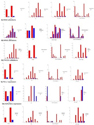
KRAS mutations were most commonly detected in the 717 CRC patients. 185 of the 397 tested patients (46.60%) had one or more of the 14 different KRAS mutation. 2 patients (1.08%) had ≤29 years, 13 patients (7.03%) had 30-39 years, 19 patients (10.27%) had 40-49 years, 42 patients (22.70%) had 50-59 years, 66 patients (35.68%) had 60-69 years, 42 patients (22.70%) had 70-79 years and 5 patients (2.70%) had ≥80 years (Supplementary material, Table SI-II). 79 of the patients with KRAS mutations were female (42.70%) and 106 were male (52.30%). 5 patients had stage I CRC (2.70%), 24 patients were staged with CRC stage II (12.97%), 67 patients were staged with CRC stage III (36.22%), 34 patients were staged with CRC stage IV (18.38%), while 55 patients (29.73%) were not staged at the time of the analyses. 51 of the patients with KRAS mutations (27.57%) had colon adenocarcinoma without specifying the correct location of the tumor tissue; 15 patients (8.11%) had cancer on caecum, 16 patients (8.65%) on sigmoid colon, 3 patients (1.62%) on ascending colon, 3 patients (1.62%) on transverse colon, 5 patients (2.70%) on descending colon, 65 patients (35.14%) on rectum, 3 patients (1.62%) on colon flexuras, 13 patients (7.03%) on rectosigmoid junction, while 11 patients (5.95%) had other diagnoses, i.e. cancer on other sites (Figure 1).
Most prominent were the KRAS mutations on codon 12, including G12D in 26.09%, G12V in 16.85%, G12C in 7.07%, G12A in 4.35%, G12S in 2.72% and G12R in 1.09% of the KRAS (+) patients. Frequently detected was also the typical codon 13 mutation, G13D in 22 (11.96%) and A146T in 10 (5.44%) of the KRAS (+) patients. Less frequently detected were the codon 61 mutations, Q61R, Q61H1 and Q16K, detected in total 11 patients (5.98%). Two concurent KRAS mutations were detected in 13 patients (7.07% of the KRAS (+) patients), of which 7 had two mutations on same codon, 4 on codon 12 and 3 on codon 146. Statisical analyses showed that there is no statistically significant correlation between the presence of KRAS mutations and patients’ gender (χ2 (2, N=397) = 1.3678, p=0.50465), age (χ2 (6, N=397) = 9.5788, p=0.14355), cancer stage (χ2 (4, N=397) = 4.2835, p=0.36900), and cancer site/diagnosis (χ2 (9, N = 397) = 11.2873, p=0.25653) (Supplementary material, Table SI-II).
Of the 402 patients tested for V600E BRAF mutation, only 27 patients (6.72%) tested positive; 18 men and 7 women, on average 61.32 years old. 16 have colon adenocarcinoma, seven rectal adenocarcinoma, one patient has rectosigmoidal adenocarcinoma and one patient with other diagnosis (Figure 1). Patients’ gender (χ2 (2, N=402) = 0.7570, p=0.68490), age (χ2 (6, N=402) = 8.8861, p=0.18009), cancer site/diagnosis (χ2 (9, N=402) = 6.8818, p=0.64942) and cancer stage (χ2 (4, N=402) = 4.0182, p=0.40355), were not significantly correlated to the presence of BRAF mutations (Supplementary material, Table SI-II).
Of the 345 patients tested for PIK3CA alterations, 31 patients (8.99%) had one or more alterations; 12 men (38.71%) and 19 women (62.29%), with mean age 61.90 years. 13 patients (41.94%) had colon adenocarcinoma with unspecified location, 8 patients (25.81%) had rectal adenocarcinoma, 4 patients (12.90%) had adenocarcinoma of caecum, 2 patients (6.45%) had adenocarcinoma on flexuras, 2 (6.45%) on other sites, and per one patient on sigmoid colon and transverse colon (3.23%) (Figure 1). 9 of these patients had E542K mutation, 11 patients had E545K mutation, 8 patients had H1047R mutation and one patient had two mutations E545K and H1047R. Sum up of the results from KRAS, BRAF and PIK3CA mutations results in 155 triple negative patients (21.62% of total included patients). Gender was shown to be significantly correlated to the presence of PIK3CA alteraitons (χ2 (2, N=345) = 6.3278, p=0.04226) and cancer site/diagnosis (χ2 (9, N=345) = 18.0269, p=0.03486), but not significantly correlated to patients’ age (χ2 (6, N=345) = 2.2940, p=0.89077) and cancer stage (χ2 (4, N=345) = 3.3648, p=0.49874). More detailed statistics are available in Supplementary material (Table SI-II).
Regarding the other genes, of the 151 patients tested for NRAS alterations, only 8 (5.30%) tested positive (4 men and 4 women): 6 patients exhibited Q61X mutation, and 2 patients exhibited G12X mutation. 2 of them were aged 40-49 years, 3 were 50-59 years old, 1 patient was 60-69 years old, and 2 patients were 70-79 years old. 3 patients have colon adenocarcinoma without specified location, one has cancer on sigmoid colon, 3 patients had rectal cancer and one patient had cancer on rectosigmoid junction. Five patients were not staged at the time of analyses, while one patient had stage II cancer, and two patients had stage III cancer (Figure 1). None of the clinicopathological factors was not significantly correlated to the observed NRAS mutations (Supplementary material, Table SI-II).
In only one of the 13 tested patients, an exon 19 EGFR deletion was detected in 74 years-old man with colon adenocarcinoma with pleural metastasis, without other genetic alterations. Also, exon 20 insertion was detected in 64 years-old man with planocellular colon cancer. For both patients, no information was available regarding the cancer stage at the time of the analyses. More detailed data representations are given in Supplementary material (Table SI-II). Furthermore, no ALK rearrangements were found in the 7 tested patients. PD-L1 was expressed in half of the 8 tested patients (n=4), three men and one woman; three of them had colon adenocarcinoma with metastases and one patient with hyperplastic colon polyps and sigmoid adenocarcinoma; all four patients had stage IV cancers. Patients’ gender is not significantly correlated with PD-L1 expression (χ2 (1, N=8) = 0.5333, p=0.46521), cancer site/diagnosis (χ2 (1, N=8) = 2.00000, p=0.15730), and cancer stage (χ2 (1, N=8) = 1.1429, p=0.28505), but was significantly associated with patients’ age (χ2 (3, N=8) = 8.0000, p=0.04601) (Supplementary material, Table SI-II). HER2/NEU was expressed 3 of the 28 tested patients (10.71%), all of which men with colon adenocarcinoma. Patients’ age (χ2 (1, N=28) = 0.9164, p=0.33843), gender (χ2 (5, N=28) = 4.7040, p=0.45306) and cancer stage (χ2 (3, N=28) = 1.1743, p=0.75971) were not significantly correlated to the HER2/NEU expression, but the cancer site/diagnsosis was (χ2 61, N=28) = 13.1911, p=0.04010) (Supplementary material, Table SI-II). Finally, 13 patients (1.81%) were tested for DPYD mutations (11 men and two women). Patients were tested for the following mutations: DPYD*2A (c.1905+1G>A, 100%), DPYD*13 (c.1679T >G, 92.31%), DPYD D949V (c.2846A>T, 100%), DPYD IVS10 (100%) and DPYD*6 (c.2194G>A, 30.77%). Of them, only one patient was heterozygous for DPYD*6 (7.69%).
Logistic regression analysis of the clinicopathological parameters that were significantly correlated to presence of specific genetic alterations was performed, with considering the values for the variables with highest frequency as references. Of the correlated variables, male patients [OR, 0.62654; 95% CI, 0.398942.50667; p=0.04235] have low probability for absence of PIK3CA alterations when CRC is diagnosed. Regarding the cancer site/diagnosis and PIK3CA alterations, there are higher probabilities that patients with adenocarcinoma on sigmoid colon [OR, 35.00000; 95% CI, 4.79504 255.47239; p=0.00046], rectal adenocarcinoma [OR, 13.3750; 95% CI, 6.52080 27.43383; p=0.00000] and synchronous cancers [OR, 13.00000; 95% CI, 3.08555 54.77149; p=0.04235] will have PIK3CA alterations. Logistic regression analysis was not performed for the correlation between PD-L1 and age due to the small sample size. Regarding the correlation between HER2/NEU expression and cancer site/diagnosis, patients with adenocarcinoma on sigmoid colon, caecum, rectum, rectosigmoid junction and other cancer sites have higher probabilities of having HER2/NEU expressed (Table 2). However, the latter results should be carefully considered due to the insufficient sample size.
Concurent alterations of two or more genes, presented in details in Table 3, were observed in 36 patients (5.02%), 17 men and 19 women, with mean age of 60.56 years; 24 patients with adenocarcinoma of colon, 10 patients of rectum, one with adenocarcinoma of rectosigmoid junction and one patient with other cancer type. All 36 patients had two genes altered. Two thirds, i.e. 25 patients (69.44%) had KRAS and PIK3CA concurrent alterations; predominantly codon 12/13 alterations (G12D, G12C, G12A, G12V, G13D) with E542K (9/25, 36%), E545K (6/25, 24%), H1047R (5/25, 20%) or their combination (H1047R/E545K, 1/25). KRAS A146T and PIK3CA H1047R were found concurent in three patients (12.50%), while KRAS Q61H and PIK3CA E545K in one patient. Four patients (11.11%) had KRAS and BRAF mutually alterated, two KRAS and NRAS, two BRAF/PIK3CA (E545K) and one patient EGFR/PD-L1.
Furthermore, 610 of the 717 included CRC patients (85.08%) were tested for MSI. Of them, 56(9.18%) had microsatelite instability; 35 men (62.5%) and 21 women (37.5%), with total mean age of 60.55 (Table 4). The women with MSI (+) CRC were significantly younger than the men (57.43 years vs. 60.49 years). 8 of the 56 patients (14.28%) were MSI-low (MSI-L), and the rest 48(85.72%) MSI-high (MSI-H). More than two thirds of the MS instable patients had colon adenocarcinoma (39/56, 69.64%), followed by rectal adenocarcinoma (12/56, 21.43%), recto sigmoidal adenocarcinoma (4/56, 7.14%) and one patient with synchronous cancers. 15 of the MSI instable patients (26.79%) had one or more gene alterations; most commonly on the KRAS codon 12, solely (8/15, 53.33%) or accompanied with PIK3CA alteration (4/15, 26.67%), or exhibited BRAF V600E mutation (2/15, 13.33%). The results may be influenced by the fact that most of the patients analyzed for MSI are not paralelly analyzed for genetic alterations. Of the 56 MSI patients, 31 patients were not staged at the time of the analyses (18 men and 13 women); of the remaining 25, only one patient had stage I cancer, 10 had stage II (8 men and 2 women), 8 patients had stage III cancers (5 men and 3 women) and 6 patients had stage IV cancers (4 men and 2 women).
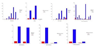
554 patients (90.82%) had microsateite stability; 339 men (61.19%) and 215 women (38.81%), with total mean age of 56.75 years. The MSS women were significantly older than the men (63.75 years vs. 61.67 years) (Table 4). 186 of the patients (33.57%) have rectal adenocarcinoma, 177(31.95%) have colon adenocarcinoma without specified cancer site, 51(9.21%) have adenocarcinoma of sigmoid colon, 46(8.30%) on other sites, 33(5.96%) on rectosigmoid junction, 28(5.05%) on caecum, 15(2.71%) on ascending colon, 9(1.62%) on transverse colon, 6(1.08%) on descending colon and 3(0.54%) on colon flexuras. 212 of the patients (38.27%) were not staged at the time of conducting analyses, 166(29.96%) had stage III cancer, 113(20.40%) had stage II cancer, 46(8.31%) had stage IV cancer, 16(2.89%) had stage I cancer and one patient was staged with stage 0 cancer (Figure 2, Supplementary material, Table SI-III).
Statistical analysis with Pearson’s Chi-square test showed that MMR protein expression was not significantly associated with patient age (χ2 (6, N=610) = 11.0926, p=0.08556), gender (χ2 (2, N=610) = 4.2407, p=0.11999), cancer diagnosis (χ2 (9, N=610) = 2.4863, p=0.18726), and cancer stage (χ2 (5, N=610) = 7.4284, p=0.19068). Also, no statistically significant association was found with the occurrence of gene alterations in KRAS (χ2 (1, N=298) = 0.1284, p=0.72005), BRAF (χ2 (6, N=305) = 0.0006, p=0.97968) and PIK3CA (χ2 (6, N=264) = 0.8355, p=0.36069) (Supplementary material, Table SI-III). The graphical representation of the MMR protein expression and clinicopathological parameters based on Pearson Chi-square test is given in Figure 2.
Table 1: General data for the patients included in the clinical study.
| Parameter |
Gender |
Age |
| Male |
Female |
(mean ± SD) |
< 29 years |
< 39 years |
40-49 years |
50-59 years |
60-69 years |
70-79 years |
≥ 80 years |
| Number |
437 |
280 |
61.80 ± 10.31 |
3 |
25 |
62 |
163 |
291 |
162 |
11 |
| Percentage |
60.95% |
39.05% |
0.42% |
3.49% |
8.65% |
22.73% |
40.59% |
22.59% |
1.53% |
| Parameter |
Tumor site |
Stage |
| Colonis |
Caecum |
Sigmoid colon |
Ascending colon |
Tra nsverse colon |
Descending colon |
Flexuras |
Rectum |
Rectosig moidal |
Other diagnoses |
0 |
I |
II |
III |
IV |
NA |
| Number |
277 |
37 |
66 |
20 |
11 |
9 |
5 |
229 |
40 |
23 |
1 |
20 |
143 |
210 |
69 |
274 |
| Percentage |
65.02% |
8.69% |
15.49% |
4.69% |
2.58% |
2.11% |
1.17% |
31.94% |
5.58% |
3.21% |
0.14% |
2.79% |
19.94% |
29.29% |
9.62% |
38.21% |
Table 2: Results of logistic regression analysis of statistically signficant varialbles.
| Variable |
Regression coefficient (B) |
OR at 95% CI (lower 95%-upper 95% confidence limit) |
Wald Z value |
p-value |
| Presence of PIK3CA alterations |
| Female |
Reference |
| Male |
0.46755 |
1.59607 (1.01627 – 2.50667) |
0.01614 |
0.91896 |
| Absence of PIK3CA alterations |
| Female |
Reference |
| Male |
-0.46755 |
0.62654 (0.39894 – 0.98399) |
-2.030 |
0.04235 |
| Presence of PIK3CA alterations |
| Colon (unspecified) |
Reference |
| Caecum |
-0.75726 |
0.46895 (0.17027 – 1.29156) |
-1.465 |
0.14292 |
| Sigmoid colon |
3.55535 |
35.000000 (4.79504 – 255.47239) |
3.506 |
0.00046 |
| Ascending colon |
14.20289 |
10 000 + |
0.039 |
0.96904 |
| Transverse colon |
1.38629 |
4.0000 (0.44708 – 35.78757) |
1.240 |
0.21500 |
| Descending colon |
14.20289 |
10 000+ |
0.031 |
0.97530 |
| Rectum |
2.59339 |
13.3750 (6.52080 – 27.43383) |
7.075 |
0.0000 |
| Flexuras |
0.40547 |
1.5000 (0.25064 – 8.97694) |
0.444 |
0.65692 |
| Rectosigmoid junction |
14.20289 |
10 000 + |
0.056 |
0.95525 |
| Other cancer sites/diagnoses |
2.56495 |
13.00000 (3.08555 – 54.77149) |
3.495 |
0.00047 |
| Absence of PIK3CA alterations |
| Colon (unspecified) |
Reference |
| Caecum |
0.75726 |
2.13243 (0.77426 – 5.87304) |
1.465 |
0.14292 |
| sigmoid colon |
-3.55535 |
0.02857 (0.00391 – 0.20855) |
-3.506 |
0.00046 |
| Ascending colon |
-14.202893 |
0.0000 (0.0000 – 10 000+) |
-0.039 |
0.96904 |
| Transverse colon |
-1.38629 |
0.25000 (0.02794 – 2.23672) |
-1.240 |
0.21500 |
| Descending colon |
-14.202894 |
0.00000 (0.00000 – 10 000+) |
-0.031 |
0.97530 |
| Rectum |
-2.593390 |
0.07477 (0.03645 – 0.15336) |
-7.075 |
0.00000 |
| Flexuras |
-0.40547 |
0.66667 (0.11140 – 3.98975) |
-0.444 |
0.65692 |
| Rectosigmoid junction |
-14.20289 |
0.00000 (0.00000 – 10 000+) |
-0.056 |
0.95525 |
| Other cancer sites/diagnoses |
-2.56495 |
0.07692(0.018260 – 0.32409) |
-3.495 |
0.00047 |
| Presence of HER2/NEU expression |
| Colon (unspecified) |
Reference |
| Caecum |
13.082630 |
10 000 + |
21.411 |
0.00000 |
| Sigmoid colon |
-15.202890 |
0.0000 |
-3.422.364 |
0.00000 |
| Ascending colon |
0.00000 |
1.00000 (0.25010 – 3.99844) |
0.000 |
1.00000 |
| Ttransverse colon |
0.00000 |
- |
- |
- |
| Descending colon |
0.00000 |
- |
- |
- |
| Rectum |
15.202890 |
10 000 + |
5.930 |
0.00000 |
| Flexuras |
0.00000 |
|
|
|
| Rectosigmoid junction |
15.20289 |
10 0000 + |
21511.860 |
0.00000 |
| Other cancer sites/diagnoses |
15.20289 |
10 000 + |
21511.860 |
0.00000 |
Table 3: Descriptive data of patients with concurent genetic alterations.
| Patient |
Diagnosis |
stage |
Gender |
Age |
Alteration 1 |
Alteration 2 |
| P1 |
Adenocarcinoma colonis transversi |
IIIB |
F |
54 |
KRAS G12D |
PIK3CA E542K |
| P2 |
Adenocarcinoma coloni |
IIIB |
F |
66 |
KRAS G12D |
PIK3CA E545K |
| P3 |
Adenocarcinoma recti |
IVB |
M |
58 |
KRAS G12C |
PIK3CA E545K |
| P4 |
Adenocarcinoma recti |
I |
M |
56 |
KRAS G12A |
BRAF V600E |
| P5 |
Adenocarcinoma colonis flexurae lienalis |
IVA |
F |
66 |
KRAS G13D |
PIK3CA H1047R |
| Patient |
Diagnosis |
stage |
Gender |
Age |
Alteration 1 |
Alteration 2 |
| P6 |
Adenocarcinoma colonis |
NA |
F |
38 |
KRAS G12D |
PIK3CA E545K |
| P7 |
Adenocarcinoma coecum |
IVB |
M |
54 |
KRAS G12A |
PIK3CA H1047R |
| P8 |
Signed ring cell carcinoma of the colon (hepatic flexurae) |
IIB |
F |
71 |
KRAS A146T |
PIK3CA H1047R |
| P9 |
Adenocarcinoma recti |
IIB |
F |
57 |
KRAS G12D, G12C |
PIK3CA E545K |
| P10 |
Adenocarcinoma rectI metastaticum hepatis |
IV |
F |
66 |
KRAS G12V |
PIK3CA 545K, H1047R |
| P11 |
Adenocarcinoma colonis |
IIA |
M |
52 |
KRAS A146T |
PIK3CA H1047R |
| P12 |
Adenocarcinoma recti |
NA |
M |
65 |
KRAS G12C, G13D |
PIK3CA E542K |
| P13 |
Adenocarcinoma colonis |
IV |
F |
66 |
KRAS G12V |
PIK3CA E542K |
| P14 |
Adenocarcinoma colonis |
NA |
M |
64 |
KRAS G12C |
PIK3CA E542K |
| P15 |
Adenocarcinoma recti |
NA |
F |
38 |
KRAS G12D |
PIK3CA E542K |
| P16 |
Adenocarcinoma recti |
NA |
M |
70 |
KRAS G12D |
NRAS Q61X |
| P17 |
Adenocarcinoma recti |
NA |
M |
63 |
KRAS G12D |
BRAF V600E |
| P18 |
Adenocarcinoma colonis sygmoidalys |
NA |
F |
67 |
BRAF V600E |
PIK3CA E545K |
| P19 |
Adenocarcinoma colonis |
IIIC |
M |
70 |
BRAF V600E |
PIK3CA E545K |
| P20 |
Adenocarcinoma recti |
I |
M |
51 |
KRAS G12S, A146T |
BRAF V600E |
| P21 |
Ca. planocellularae metastaticum reg coli lat sin |
NA |
M |
64 |
EGFR exon20ins |
(+)-PD-L1 |
| P22 |
Adenocarcinoma colonis |
NA |
F |
46 |
KRAS G12D |
NRAS G12X |
| P23 |
Adenocarcinoma rectosigmae ms pulmonum |
NA |
M |
71 |
KRAS G12D |
DYDP |
| P24 |
Adenocarcinoma colonis |
IIIB |
M |
51 |
KRAS G13D |
PIK3CA H1047R |
| P25 |
Adenocarcinoma colonis |
NA |
F |
74 |
KRAS G12D |
PIK3CA E542K |
| P26 |
Adenocarcinoma rectosigmae |
IIC |
M |
42 |
KRAS codon 12/13 |
DYDP*6 |
| P27 |
Adenocarcinoma colonis |
IIIB |
F |
64 |
KRAS G12D |
PIK3CA H1047R |
| P28 |
Adenocarcinoma colonis |
IVA |
F |
42 |
KRAS G12D |
PIK3CA E542K |
| P29 |
Adenocarcinoma colomis |
IIIC |
F |
70 |
KRAS G12D |
PIK3CA E545K |
| P30 |
Adenocarcinoma coecci |
II |
F |
73 |
KRAS G13D |
PIK3CA E542K |
| P31 |
Adenocarcinoma recti |
NA |
M |
71 |
KRAS G12C |
PIK3CA H1047R |
| P32 |
Adenocarcinoma coecci |
IIA |
M |
66 |
KRAS Q61H |
PIK3CA E545K |
| P33 |
Adenocarcinoma intestinii crassi et sessile serrated adenoma |
NA |
F |
62 |
KRAS G12V |
BRAF V600E |
| P34 |
Adenocarcinoma coecci |
NA |
F |
80 |
KRAS A146T |
PIK3CA H1047R |
| P35 |
Adenocarcinoma coecci |
IIIC |
F |
48 |
KRAS G12C, G12V |
PIK3CA E545K |
| P36 |
Adenocarcinoma intestini crassii |
NA |
M |
64 |
KRAS G12V |
PIK3CA E542K |
Table 4: Descriptive information of examined patients for MMR protein expression.
| MSI result |
Number of patients (%) |
Mean age (years)
| Diagnosis of the patients |
| Adenocarcinoma colonis |
Adenocarcinoma recti |
Adenocarcinoma rectosigmoidalis |
Other diagnoses |
| MSI |
Total |
56 |
9.18% (of total number of patients) |
60.55 |
39 |
12 |
4 |
1 |
| M |
35 |
62.5% |
60.49 |
21 |
9 |
4 |
1 |
| F |
21 |
37.5% |
57.43 |
18 |
3 |
0 |
0 |
| MSS |
Total |
554 |
90.82% (of total number of patients) |
56.75 |
204 |
8 |
7 |
335 |
| M |
339 |
61.19% |
61.67 |
117 |
5 |
6 |
211 |
| F |
215 |
38.81% |
63.78 |
87 |
3 |
1 |
124 |
Discussion
The response of CRC to current therapeutic approaches is highly variable, reflecteing the elevated heterogeneity of the disease. This, together with an increased availability of tageted therapeutic approaches, stresses the need for more comprehensive molecular characterization of each CRC sample, in order to push forward the real achievement of personalized interventions. Despite the knowledge of the significant impact of the extended molecular characterization on their clinical management, very little of it has entered the clinical routine, yet.
Here, we report an extensive molecular profiling of 717 CRC patients, that obtained results providing greast opportunities to direct more informed therapeutic decisions. Overall, 225 of the tissue samples (31.38%) had at least one somatic mutation, of which 36(5.02%) had two mutations. The somatic variation frequency in CRC detected in this study is lower than in other multigene panel sequencing studies (e.g. 98.46% in [18], 97.55% in [19], 81.8% in [20], 76.41% in [21], 69% in [7], with average of three variants per sample; 68.84% in [22]). Still, a strict conclusion that frequency of gene mutations in North Macedonian CRC patients cannot be drawn, since it may be result of the study design – as a retrospective study.
KRAS and NRAS mutations
KRAS and NRAS are members of the RAS gene family. KRAS is one of the most frequently mutated genes in CRC, with alterations observed in approximately 37.8 – 51.4% of the CRC patients [14,19–25], although lower overall frequencies (9-17%) are also reported [7,18,26]. Mutations are concentrated in exon 2, codons 12 (G12D, G12V, G12C 70-80%) and codon 13 (Q61H, Q61R 15-20%). Also detected are mutations in exon 3, codons 59-61 and exon 4, codons 117 and 146 (A146T and A146V). It is established that KRAS and NRAS exon 3 mutations occur more frequently as result of acquired resistance to EGFR-antibody therapy than in de novo setting [27]. In the present study, KRAS (codon 12, 13 and 61) mutations occurred in 46.60% of the tested patients, of which 37.78% on codons 12 and/or 13, 3.53% on codon 146 and 3.02% on codon 61. Concerning single mutation frequencies, our study showed in some cases comparable (G12C: 7.07% vs 8.5%; G13D: 11.96% vs 14.1%; Q61H: 2.16% vs 1.40%; Q61K: 0.54% vs 1.40%; A146T: 5.44% vs 4.20%), lower (G12S 2.72% vs 9.9%; G12D: 26.09% vs 32.4%) or higher frequencies (G12A: 4.35% vs 2.8%; G12V: 16.85% vs 11.30%) that other mutligene sequencing studies [28]. These variations, such as G12C, G12V, G13D, Q61H and A146T, G12D and G13D are previously reported in many CRC multigene sequencing studies and described as pathogenic for the KRAS gene and recorded in the COSMIC database [7,18,22].
NRAS variations occured in 5.30% of the tested patients, which is in line with available studies (3.0-6.0%) [14,18,20–24,26] and may limit efficacy of panitumumab, especially in wt KRAS tumors [24]. Mutations most frequently affect codonс 12(78%), 13(18%) and 61(4%) [23].
Concerning gender, data has been inconclusive. Some studies report equal distribution of RAS mutations (43.8% in men vs. 43.3% in women, p=0.006) [14], while other study indicates higher prevalence in women than in men (73.4% vs. 66.2%) [28]. In our study, RAS mutations slightly predominate in women (29.64% vs. 25.17%), although no statistically significant correlation was found between presence of KRAS/NRAS mutations and gender, which was also the case in [28]. In terms of tumor location, findings are incosistent: some studies indicate that KRAS mutation does not correlate with tumor location, while others indicate higher occurrence of KRAS mutations and in the caecum area [14]. Here, most of the KRAS mutations occurred in patients with unspecified location of colon adenocarcinoma (27.57%) and rectal adenocarcinoma (35.13%), but cancer site was not significantly asscoiated with presence of KRAS/NRAS mutations (p=0.7977). Cosnidering tumor site, KRAS and NRAS mutations have similar clinical and pathological features. One study indicates association of KRAS mutations with tumor location but only in men [28]. Further, 10 of the KRAS mutated patients (5.41%) had mCRC; five of which had liver metastases, and one patient per each of remaning locations: omenti majors, lungs, peritoneum, lymph nodes and ovarii; although other studies report high indicence of lung metastases in KRAS mutated patients [23]. Regarding location of NRAS tumors, they were identified on the proximal colon, distal colon and rectum in 33%, 36% and 31% of the cases, respectively [23].
KRAS and NRAS mutations on exons 2, 3 and 4 have exceptional clinical significance, as they indicate lack of anti-EGFR antibody efficacy, with worse PFS and OS. Previous meta-analyisis indicate that ~53% of CRC tumors (~42% with KRAS exon 2 mutations and ~11% with KRAS exon 3 or 4 or NRAS exon 2, 3 or 4 mutations) are likely resitant to anti-EGFR antibodies [29]. In particular, the presence of a KRAS mutation is associated with a downstream activation of the Ras/MAPK pathway, leading to cell proliferation that cannot be significantly inhibited by cetuximab that acts upstream of the K-ras protein. Also, KRAS mutations are found to be associated with resistance to EGFR kinase inhibitors gefitinib and erlotinib [30].
Evaluation of single KRAS mutations reveal that no individual mutant KRAS allele is consistently associated with PFS or OS outcomes, although some studies sugges that CRC tumors with KRAS G13D mutations have favorable outcomes versus patients with other KRAS mutations [24]. On the other hand, the G12V mutation has been reported to be more aggressive that other KRAS mutations. However, no influence of the mutation type (i.e. codon 12 v codon 13 or G12V mutation vs. other mutations) on response rate or prognosis is detected [31].
EGFR, BRAF and PIK3CA mutations
In the present study, EGFR variations were detected in 0.28% of the total patients, which is lower than the seldomly reported mutation frequency in other studies (e.g. 1.72% [20], 1.2% [21]). We detected exon 19 deletion and exon 20 insertion in one instances each.
Furthermore, 6.72% of the tested patients had BRAF V600E mutation, which is in line with available scientific data (3.23 – 9.60% [7,20,21,24,26]. 81.48% of the patients with mutated BRAF had exclusively BRAF mutations, possibly representing the best subset for a target treatment with BRAF inhibitors alone or combined with anti-EGFR agents. 18.52% of the patients carried other mutations on KRAS or PIK3CA that can be expected to be involved in primary resistance to anti-BRAF therapies, providing contraindications to single target approaches. We observed very low incidence of MSI patients with BRAF mutations (3.57% of all MSI patients, i.e. 7.41% of all patients with BRAF mutations), than published in other studies, where, for example, just under one-third of BRAF-mutant tumors also had MSI, and the same proportion of tumors with MSI contained BRAF mutations [32].
BRAF mutations were more frequent in female (66% vs. 34%) [23], more frequently right-sided (64% right, 13% left, 23% rectal) also in comparison to KRAS mutated (p<0.0001) and all wt (p<0.0001) tumors. They were more frequently associated with mucinous histology in comparison to KRAS mutated (p=0.004) and all wt (p=0.005) tumors and showed a higher pathological involvement of locoregional lymph nodes in comparison to all wt (pN1–2: BRAF mutated 87% vs all wt 70%, p=0.012) [23]. They are also shown to be significantly correlated to the pathological types of cancer previously [19], but that was not the case in our study.
BRAF mutations are significant negative prognostic marker for patients with mCRC; the finding is supported by numerous randomised studies with specific chemotherapy regimens [14,30,33,34]. Evidence of BRAF mutations as a negative predictive biomarker for EGFR antibody therapy is accumulating [24], while it is clearly established that EGFR antibodies cetuximab and panitimumab are more beneficial in patients with RAS-wt/BRAF-wt tumors than in RAS-wt/BRAF-mutant tumors, which has poorer PFS [24,30,33]. Furthermore, somatic BRAF V600E mutations are associated with sporadic cases of dMMR, showing an MSI phenotype, and thus are used as differentiating tool between HNPCC from sporadic MSI-H CRC patients [35]. Also, OS for patients with BRAF mutations is lower than for patients with KRAS exon 2 mutations and wt KRAS (8.8 months vs. 14.4 and 20.1 months) [36] and OS of 10.4 months with BRAF mutations vs. 34.7 months for BRAF wt tumors [32].
In the present studies, PIK3CA variations appeared in 8.50% of the tested patients, which is in line with frequencies in other studies (6.67 – 11.30% [18,24–26]), altohugh there are many studies reporting higher frequencies (15.00 – 25.24% [7,19–22]). Furthermore, PIK3CA alterations alone occurred rarely (n=2, 6.45%), and more frequently were accompanied by BRAF V600E (n=2, 6.45%) and KRAS mutations (n=27, 87.10%), which is a 2-fold-lower than previously reported frequency (15.3%) [21]. PIK3CA mutations often co-occur with KRAS or BRAF mutations [20–22,26,28,37], but there is insufficient evidence for their use as mutual biomarkers of resistance to EGFR-antibody therapy. Studies exist that clearly prove the need to examine the mutation status of the PI3K signaling pathway prior to treatment with EGFR receptor antagonists [13], and studies that fail to observe link between PIK3CA mutation status and cetuximab response in CRC patients (e.g. [30]). The latter may be due to the low PIK3CA mutation frequency, which was also the case in the present study, further indicating the need for validation in a larger patient study.
ALK rearrangements, PD-L1 and HER2/NEU expression
ALK rearrangements did not occur in patients from our study. In fact, such alterations are very rarely reported, only in very large studies, e.g. conducted on 639 patients, with very low incidence (0.16%) [20]; or on 653 patiensts, with 0.2% incidence [21]. Regarding PD-L1 expression, only 1.25% of the patients were tested, of which 0.56% of all patients had high PD-L1 expression. This is far below the reported frequency (~5%) [8], most probably due to the very low testing frequency in our study. PD-L1 expression is shown not to be associated with gender, age TNM stage, tumor size and presence of lymph node metastasis, but it is associated with cancer location and differentiation, such that poor differentiation and right colon location are PD-L1 expression positive [38]. Our study, however, did include investigation of such associations due to the low sample size. Finally, HER2/NEU amplifications were found in 10% of the tested patients (0.42%, n=717), all in proximal colon. The frequency is higher than the previously reported frequencies, e.g. 5.1% all in rectosigmoid colon [28]; ~0.63% [20,21,25]. Although there are studies indicating the association of HER2 expression and CRC tumor location [43], results of our study are not conclusive due to the small sample size.
DPYD mutations
Our study revealed only one patient with single DPYD mutation – DYPD*6, that was 52-year old male with adenocarcinoma intestinii crasii. This mutation is found not to affect DPD activity in a clinically relevant manner [10]. DYPD*6 is also found to be significantly associated with adverse reactions of fluoropyrimidines in multiple studies, as the one of Del Re et al. [39]; the secondary analysis of the PanEuropean Trials in Alimentary Tract Cancer study, that provided evidence of the association of c.2194G>A variant with clinically-relevant ADRs in FOLFOX4-treated patients (5FU, leucovorin, oxaliplatin) [40]; the TOSCA randomized trial that enrolled colon cancer patients given 3 or 6 months of either FOLFOX-4 or XELOX adjuvant chemotherapy [41]. Boige et al.’s cohort confirmed association of DPYD*6 with hematological adverse events, neutropenia [40] and leukopenia [41] in CRC patients, they are also studies that indicate the lack of such association with fluoropyrimidine toxicity [42,43]. Finally, current guidelines of 5-FU dose adjustment [42] do not include recommendations for DPYPD*6 mutationfs.
Concurrent genetic alterations
In our samples, concurrent alterations of two genes were observed in 36 patients (5.02%). The frequency of concurrent mutations is somewhat lower than in other studies (e.g. 7.55% [26]). Here, 3.35% of all patients had KRAS/PIK3CA variations, predominantly codon 12/13 alterations (G12D, G12C, G12A, G12V, G13D) with E542K (8/24, 33.33%), E545K (6/24, 25.00%), H1047R (4/24, 16.67%) or their combination (H1047R/E545K, 1/24). The frequency is somewhat different than available data, as other studies report concurrent KRAS/PIK3CA mutations in 20.29% of the patients, of which 48% with codon 12 and 8% with codon 13 alterations [22]. This frequent co-occurrence leads to constitutive activation of two different pathways downstream of EGFR, which, also for PIK3CA/BRAF mutations, may be the reason of scant success of PI3K inhibitors in mCRC [20].
Four patients (11.11%) had KRAS and BRAF mutually alterated, two KRAS and NRAS (5.56%), two BRAF/PIK3CA (E545K) (5.56%), and one patient EGFR/PD-L1. Large studies report rare co-occuring gene alterations, such as KRAS/NRAS variations in 0.94% and KRAS/BRAF variations in 0.63% of the patients [24]. Morevoer, KRAS and NRAS mutations are known to be mutually almost exclusive [21], which was also confirmed with our study. Additionally, 25 patients (69.44%) had KRAS and PIK3CA concurent alterations; predominantly codon 12/13 alterations (G12D, G12C, G12A, G12V, G13D) with E542K (9/25, 36%), E545K (6/25, 24%), H1047R (5/25, 20%) or their combination (H1047R/E545K, 1/25). KRAS A146T and PIK3CA H1047R were found concurent in three patients (12.50%), while KRAS Q61H and PIK3CA E545K in one patient.
This is partially in line with scientific data, as it indicates that mutations in KRAS, BRAF, NRAS [20], and also PIK3CA appeared to be mutually exclusive [18], while other studies indicate that BRAF mutations are almost exclusively non-overlapping with RAS mutations, but coexist with PIK3CA mutations in 1.8-13% of the patients [14,26,32]. Such combined mutations in NRAS, BRAF and PIK3CA predict resistance to anti-EGFR antibodies with a statistically significant value [18]. Considering clinical significance, combined NRAS, BRAF and PIK3CA mutation frequency was significantly correlated to anti-EGFR resistance phenotype [18], but we could not investigate this in our study due to the lack of information regarding the therapeutic regimens of the tested patients.
MSI
MSI is a DNA abnormality that occurrs in about 10-20% of sporadic CRC patients, 3.5% in metastatic CRC and 95% od inherited CRCs, that have Lynch syndrome, HNPCC and FPS [6,14,34]. MSI tumors have distinct features, such as origin in the proximal colon, prominent lymphocytic infiltrate, poorly differentiated morphology, mucinous or signet ring differentiation and association with a favorable prognosis in early-stage CRC. This supports that dMMR tumors have reduced metastatic potential [34].
In our study, of the 610 tested CRC patients, 9.18% (n=56) were MSI, of which 8(14.28%) were MSI-L and the remaining 48(85.72%) were MSI-H. This is in line with other studies (e.g. 20%, half of which MSI-H [25]; or where 65% were MSS, 10% were MSI-L and 25% were MSI-H [44]). MSI-H comprise ~15% of CRCs, are frequently poorly differentiated with mucinous histological features with intraepithelial lymphocytic infiltration [45,46], with periand intratumoral lymphocytic invasion and good prognosis [45]. They are seen in female patients, arise in proximal colon, as less aggressive, present at lower stage and have increased responsiveness to certain adjuvant chemotherapy [46]. In fact, MSI is known to be more common among stage II (~20%) than III (~12%) and is even less frequent in stage IV CRC (~4%) [47].
Considering age, a recent retrospective analysis demonstrated that the median age of MSI patients is 67 years, with more frequent poor differentiation (58%), and that 45% of the patients with MSI phenotype had stage IV disease at presentation [45]; also, mean age of 72 is reported [32]. In our study, MSI patients were younger on average (60.55 years), but, it should be considered that most patients (53.57%) were not staged at the present time, and therefore, appropriate conclusion cannot be drawn. Also, more than two thirds of the MSI patients had colon adenocarcinoma (39/56, 69.64%), followed by rectal adenocarcinoma (12/56, 21.43%), recto sigmoidal adenocarcinoma (4/56, 7.14%) and one patient with synchronous cancers. This frequency is in accordance with data from previous studies. In other study, MSI was significantly associated with tumor site, side of colon with cancer and histological CRC type, but was not significantly associated with patient age, gender or ethnic group [46]. Our statistical analysis did not reveal such associations.
Loss of DNA MMR activitiy accelerates the rate of accumulation of mutations in other genes involved in apoptosis and growth control, especially in MSI-H patients [25], as for example, one study discovering 14.26% of the MSI patients to have ≥20 mutations [37]. Most common are BRAF mutations, that indicate a sporadic origin and essentially exclude diagnosis of Lynch syndrome [34]. In our study, 26.79% of the MSI instable patients had one or more gene alterations; most commonly on the KRAS codon 12 and 13, solely (53.33%) or accompanied with PIK3CA alteration (26.67%), or exhibited BRAF V600E mutation (13.33%), which is in line with previous studies (KRAS 54%, BRAF 21%, PIK3CA 32%) [37]. In our study, BRAF V600E mutations were present in 30% of patients with MSI [45], similar to other studies [14,27,35], as well as in 18 MSS patients (3.25%), which is in line with previous studies (4-12% [14,35]). Such higher frequency of BRAF mutations in MSI tumors than in MSS tumors is also confirmed in a pool analysis of four phase III studies (CAIRO, CAIRO2, COIN and FOCUS). The same pooled analysis showed PFS and OS to be significantly worse for patients with tumours with MSI when compared with those with tumours showing MSS and for patients with BRAF-mutant tumours when compared with those with BRAF wild-type tumours. However, there is no significant survival difference in BRAF-mutant patients with MSI and MSS s (PFS: 6.3 versus 7.8 months, respectively; OS: 15.0 versus 17.3 months, respectively; the same is true for MSI patients with and without BRAF mutations (PFS: 6.1 versus 6.3 months, respectively; OS: 11.7 versus 15.0 months, respectively) [34]. According to present data, analysis for BRAF V600E mutation is a highly specific molecular marker to exclude HNPCC and should be used in parallel with quantitative dMMR methylation analysis.
Conclusion
The data from the current study confirm that CRCs consists of group of heterogenous disorders with a large number of diverse sets of genetic changes in oncogenes and tumor suppresor genes. Our study was able to generate robust and comprehensive genetic information, refining the identification of reliable and reproducible biomarkers of response/resistance to the targeted treatment of CRC. As, such, it is the first to provide such detailed genetic profiling of CRC patients from North Macedonia to date, and as such, provides valuable knowledge core for further, more thorough studies. Additionally, our data straighten the view that simple distinction of tumors in RAS, BRAF or PIK3CA does not apply to CRC with combined genetic changes.
1) However, the results of this study have to be taken into consideration together with its significant limSmall sample size of patgients for certain gene alterations (e.g. EGFR, ALK, PD-L1, HER2/NEU). This limits the interpretation of subgroup analyses as type 2 errors are possible from comparing rare endpoints. Therefore, prospective large-scale studies are needed to adress this issue;
2) Lack of knowledge regarding other diseases and medications, that could influence the CRC and efficacy of therapeutic regimens;
3) Lack of knowledge regarding applied treatment regimens;
4) Lack of patients’ follow-up;
5) In terms of DPYD alterations, lack of evaluation of the effect of reactive versus preemptive DPYD genotyping, as there is no information regarding the administered therapy at the time of the analyses.
Finally, it should also be noted that the mutations were not confirmed using a second analytical technique. Further studies should be based on multigenome sequencing of CRC patients, incorporated as routine CRC patient management outside of a clinical trial setting; as recent studies suceeded to discover multitude of new somatic variants in CRC patients [7]. Such advancements in molecular profiling of CRC patients would facilitate the ability to direct treatments to the biologic features of the tumor for specific patient subsets. It will allow treatment selection towards increasing the benefits and decreasing the toxicity from ineffective therapies.
Abbreviations
5-FU: 5-Fluorouracil; ALK: Anaplastic Lymphoma Kinase; CRC: Colorectal Cancer; dMMR: DNA Mismatch Repair Deficiency; DPYD: Dihydropiyrimidine Dehydrogenase Encoding-Gene; EGFR: Epidermal Growth Factor Receptor; ESMO: European Society for Medical Oncology; FFPE: Formalin-Fixed Paraffin-Embedded; HER2: Human Epidermal Growth Factor Receptor 2; HNPCC: Hereditary Non-Polyposis Colorectal Cancer; IARC: International Agency for Research on Cancer; IHC: Immunohistochemistry; KRAS: Kirsten Rat Sarcoma Virla Oncogene; MAPK: Mitogenactivated Protein Kinase; mCRC: Metastatic Colorectal Cancer; MSI: Microsatellite Instability; MSI-H: MSI High; MSI-L: MSI Low; NA: Not Applicable; NCCN: The National Comprehensive Cancer Network; OS: Overall Survival; PD-L1: Programmed Death-Ligand 1; PFS: Progression-Free Survival; PI3K: Phosphoinositide 3-Kinase; PIK3CA: Phosphatidylinositol-4,5-Bisphosphate 3-Kinase Catalytic Subunit Alpha; RR: Response Rate; RT-PCR: Real-Time Polymerase Chain Reaction; WHO: World Health Organization.
References
- Sung H, Ferlay J, Siegel RL, Laversanne M, Soerjomataram I, Jemal A, et al. Global Cancer Statistics 2022: GLOBOCAN Estimates of Incidence and Mortality Worldwide for 36 Cancers in 185 Countries. CA Cancer J Clin. 2024; 71: 209-249.
- Siegel RL, Miller KD, Goding Sauer A, Fedewa SA, Butterly LF, Anderson JC, et al. Colorectal cancer statistics, 2020. CA Cancer J Clin. 2020; 70: 145-164.
- Ferlay J, Ervik M, Lam F, Colombet M, Mery L, Piñeros M, et al. Global Cancer Observatory: Cancer Today. International Agency for Research on Cancer, Lyon, France. 2020. Available from: https:// gco.iarc.fr/today (accessed February 9, 2022).
- Biller LH, Schrag D. Diagnosis and Treatment of Metastatic Colorectal Cancer: A Review. J Am Med Assoc. 2021; 325: 669-685.
- Muratore A, Zorzi D, Bouzari H, Amisano M, Massucco P, Sperti E, et al. Asymptomatic Colorectal Cancer with Un-Resectable Liver Metastases: Immediate Colorectal Resection or Up-Front Systemic Chemotherapy?. Ann Surg Oncol. 2007; 14: 766-770.
- Fearon ER. Molecular Genetics of Colorectal Cancer. Annu Rev Pathol Mech Dis. 2011; 6: 479-507.
- Youssef ASE-D, Abdel-Fattah MA, Lotfy MM, Nassar A, Abouelhoda M, Touny AO, et al. Multigene Panel Sequencing Reveals CancerSpecific and Common Somatic Mutations in Colorectal Cancer Patients: An Egyptian Experience. Curr Issues Mol Biol. 2022; 44: 1332-1352.
- Lee LH, Cavalcanti MS, Segal NH, Hechtman JF, Weiser MR, Smith JJ, et al. Patterns and prognostic relevance of PD-1 and PD-L1 expression in colorectal carcinoma. Mod Pathol. 2016; 29: 1433-1442.
- Pagani F, Randon G, Guarini V, Raimondi A, Prisciandaro M, Lobefaro R, et al. The Landscape of Actionable Gene Fusions in Colorectal Cancer. Int J Mol Sci. 2019; 20: 5319.
- Amstutz U, Henricks LM, Offer SM, Barbarino J, Schellens JHM, Swen JJ, et al. Clinical Pharmacogenetics Implementation Consortium (CPIC) Guideline for Dihydropyrimidine Dehydrogenase Genotype and Fluoropyrimidine Dosing: 2017 Update. Clin Pharmacol Ther. 2018; 103: 210-216.
- Morel A, Boisdron-Celle M, Fey L, Soulie P, Craipeau MC, Traore S, et al. Clinical relevance of different dihydropyrimidine dehydrogenase gene single nucleotide polymorphisms on 5-fluorouracil tolerance. Mol Cancer Ther. 2006; 5: 2895-2904.
- Cathomas G. PIK3CA in Colorectal Cancer. Front Oncol. 2014; 4: 35.
- Jhawer M, Goel S, Wilson AJ, Montagna C, Ling YH, Byun DS, et al. PIK3CA mutation / PTEN expression status predicts response of colon cancer cells to the EGFR inhibitor cetuximab. Cancer Res. 2008; 68: 1953-1961.
- Afrăsânie VA, Marinca MV, Alexa-Stratulat T, Gafton B, Păduraru M, Adavidoaiei AM, et al. KRAS, NRAS, BRAF, HER2 and microsatellite instability in metastatic colorectal cancer – practical implications for the clinician. Radiol Oncol. 2019; 53: 265-274.
- Ivanova M, Venetis K, Guerini-Rocco E, Bottiglieri L, Mastropasqua MG, Garrone O, et al. HER2 in Metastatic Colorectal Cancer: Pathology, Somatic Alterations, and Perspectives for Novel Therapeutic Schemes. Life. 2022; 12: 1403.
- Siena S, Sartore-Bianchi A, Marsoni S, Hurwitz HI, McCall SJ, Penault-Llorca F, et al. Targeting the human epidermal growth factor receptor 2 (HER2) oncogene in colorectal cancer. Ann Oncol. 2018; 29: 1108-1119.
- National Comprehensive Cancer Network. Colon cancer v.2.2023. 2022.
- Lupini L, Bassi C, Mlcochova J, Musa G, Russo M, Vychytilova-Faltejskova P, et al. Prediction of response to anti-EGFR antibody-based therapies by multigene sequencing in colorectal cancer patients. BMC Cancer. 2015; 15: 808.
- Zhuang Y, Wang H, Jiang D, Li Y, Feng L, Tian C, et al. Multi gene mutation signatures in colorectal cancer patients: predict for the diagnosis, pathological classification, staging and prognosis. BMC Cancer. 2021; 21: 380.
- Belardinilli F, Capalbo C, Malapelle U, Pisapia P, Raimondo D, Milanetti E, et al. Clinical Multigene Panel Sequencing Identifies Distinct Mutational Association Patterns in Metastatic Colorectal Cancer. Front Oncol. 2020; 10: 560.
- Malapelle U, Pisapia P, Sgariglia R, Vigliar E, Biglietto M, Carlomagno C, et al. Less frequently mutated genes in colorectal cancer: evidences from next-generation sequencing of 653 routine cases. J Clin Pathol. 2016; 69: 767-771.
- Gong J, Cho M, Sy M, Salgia R, Fakih M. Molecular profiling of metastatic colorectal tumors using next-generation sequencing: a single-institution experience. Oncotarget. 2017; 8: 42198-42213.
- Schirripa M, Cremolini C, Loupakis F, Morvillo M, Bergamo F, Zoratto F, et al. Role of NRAS mutations as prognostic and predictive markers in metastatic colorectal cancer. Int J Cancer. 2015; 136: 83-90.
- Peeters M, Oliner KS, Parker A, Siena S, Van Cutsem E, Huang J, et al. Massively Parallel Tumor Multigene Sequencing to Evaluate Response to Panitumumab in a Randomized Phase III Study of Metastatic Colorectal Cancer. Clin Cancer Res. 2013; 19: 1902-1912.
- Han SW, Kim HP, Shin JY, Jeong EG, Lee WC, Lee KH, et al. Targeted Sequencing of Cancer-Related Genes in Colorectal Cancer Using Next-Generation Sequencing. PLoS ONE. 2013; 8: e64271.
- Hsu HC, Thiam TK, Lu YJ, Yeh CY, Tsai WS, You JF, et al. Mutations of KRAS/NRAS/BRAF predict cetuximab resistance in metastatic colorectal cancer patients. Oncotarget. 2016; 7: 22257-22270.
- Van Cutsem E, Cervantes A, Adam R, Sobrero A, Van Krieken JH, Aderka D, et al. ESMO consensus guidelines for the management of patients with metastatic colorectal cancer. Ann Oncol. 2016; 27: 1386-1422.
- Kwak MS, Cha JM, Cho YH, Kim SH, Yoon JY, Jeon JW, et al. Clinical Predictors for KRAS Codon 13 Mutations in Patients with Colorectal Cancer. J Clin Gastroenterol. 2018; 52: 431-436.
- Sorich MJ, Wiese MD, Rowland A, Kichenadasse G, McKinnon RA, Karapetis CS. Extended RAS mutations and anti-EGFR monoclonal antibody survival benefit in metastatic colorectal cancer: a metaanalysis of randomized, controlled trials. Ann Oncol. 2015; 26: 1321.
- Lièvre A, Bachet JB, Le Corre D, Boige V, Landi B, Emile JF, et al. KRAS Mutation Status Is Predictive of Response to Cetuximab Therapy in Colorectal Cancer. Cancer Res. 2006; 66: 3992-3995.
- Lievre A, Bachet JB, Boige V, Cayre A, Lecorre D, Buc E, et al. KRAS Mutations As an Independent Prognostic Factor in Patients With Advanced Colorectal Cancer Treated With Cetuximab. J Clin Oncol. 2008; 26: 374-379.
- Tran B, Kopetz S, Tie J, Gibbs P, Jiang ZQ, Lieu CH, et al. Impact of BRAF Mutation and Microsatellite Instability on the Pattern of Metastatic Spread and Prognosis in Metastatic Colorectal Cancer. Cancer. 2011; 117: 4623-4632.
- Pietrantonio F, Petrelli F, Coinu A, Di Bartolomeo M, Borgonovo K, Maggi C, et al. Predictive role of BRAF mutations in patients with advanced colorectal cancer receiving cetuximab and panitumumab: A meta-analysis. Eur J Cancer. 2015; 51: 587-594.
- Venderbosch S, Nagtegaal ID, Maughan TS, Smith CG, Cheadle JP, Fisher D, et al. Mismatch repair status and BRAF mutation status in metastatic colorectal cancer patients: a pooled analysis of the CAIRO, CAIRO2, COIN and FOCUS studies. Clin Cancer Res. 2014; 20: 5322-5330.
- Bettstetter M, Dechant S, Ruemmele P, Grabowski M, Keller G, Holinski-Feder E, et al. Distinction of Hereditary Nonpolyposis Colorectal Cancer and Sporadic Microsatellite-Unstable Colorectal Cancer through Quantification of MLH1 Methylation by Real-time PCR. Clin Cancer Res. 2007; 13: 3221-3228.
- Maughan TS, Adams RA, Smith CG, Meade AM, Seymour MT, Wilson RH, et al. Addition of cetuximab to oxaliplatin-based first-line combination chemotherapy for treatment of advanced colorectal cancer: results of the randomised phase 3 MRC COIN trial. Lancet. 2011; 377: 2103-2114.
- Stadler ZK, Battaglin F, Middha S, Hechtman JF, Tran C, Cercek A, et al. Reliable Detection of Mismatch Repair Deficiency in Colorectal Cancers Using Mutational Load in Next-Generation Sequencing Panels. J Clin Oncol. 2016; 34: 2141-2147.
- Li Y, He M, Zhou Y, Yang C, Wei S, Bian X, et al. The Prognostic and Clinicopathological Roles of PD-L1 Expression in Colorectal Cancer: A Systematic Review and Meta-Analysis. Front Pharmacol. 2019; 10: 139.
- Del Re M, Cinieri S, Michelucci A, Salvadori S, Loupakis F, Schirripa M, et al. DPYD6 plays an important role in fluoropyrimidine toxicity in addition to DPYD2A and c.2846A>T: a comprehensive analysis in 1254 patients. Pharmacogenomics J. 2019; 19: 556-563.
- Boige V, Vincent M, Alexandre P, Tejpar S, Landolfi S, Le Malicot K, et al. DPYD Genotyping to Predict Adverse Events Following Treatment With Fluorouracil-Based Adjuvant Chemotherapy in Patients With Stage III Colon Cancer: A Secondary Analysis of the PETACC-8 Randomized Clinical Trial. JAMA Oncol. 2016; 2: 655.
- Ruzzo A, Graziano F, Galli F, Galli F, Rulli E, Lonardi S, et al. Dihydropyrimidine dehydrogenase pharmacogenetics for predicting fluoropyrimidine-related toxicity in the randomised, phase III adjuvant TOSCA trial in high-risk colon cancer patients. Br J Cancer. 2017; 117: 1269-1277.
- Wei X, Elizondo G, Sapone A, McLeod HL, Raunio H, Fernandez-Salguero P, et al. Characterization of the Human Dihydropyrimidine Dehydrogenase Gene. Genomics. 1998; 51: 391-400.
- Ridge SA, Sludden J, Wei X, Sapone A, Brown O, Hardy S, et al. Dihydropyrimidine dehydrogenase pharmacogenetics in patients with colorectal cancer. Br J Cancer. 1998; 77: 497-500.
- Ashktorab H, Mokarram P, Azimi H, Olumi H, Varma S, Nickerson ML, et al. Targeted exome sequencing reveals distinct pathogenic variants in Iranians with colorectal cancer. Oncotarget. 2016; 8: 7852-7866.
- Goldstein J, Tran B, Ensor J, Gibbs P, Wong HL, Wong SF, et al. Multicenter retrospective analysis of metastatic colorectal cancer with high-level microsatellite instability. Ann Oncol. 2014; 25: 1032-1038.
- Kaur G, Masoud A, Raihan N, Radzi M, Khamizar W, Kam LS. Mismatch repair genes expression defects and association with clinicopathological characteristics in colorectal carcinoma. Indian J Med Res. 2011; 134: 186-192.
- Koopman M, Kortman GAM, Mekenkamp L, Ligtenberg MJL, Hoogerbrugge N, Antonini NF, et al. Deficient mismatch repair system in patients with sporadic advanced colorectal cancer. Br J Cancer. 2009; 100: 266-273.