Introduction and rationale
Androgen Receptor (AR) expression was found to be related to a favorable prognosis in Breast Cancers (BC) expressing estrogen receptor. AR seems to inhibit the cellular proliferation induced by estradiol and to have a favorable prognostic value. AR might modulate the ER signaling by interfering with both genomic and non-genomic actions [5].
In vitro, in estrogen receptor – positive (ER-positive) ZR75 and MCF-7 cells, activated AR up-regulates the estrogen receptor – β (ERβ) promoter and lastly inhibits cancer cell growth, as ERβ prevalently acts as a tumor suppressor in BC [15].
In several specimens of Tamoxifen-resistant BC, low ER mRNA and high AR mRNA levels were detected. Treatment with the anti-androgen Bicalutamide reversed this resistance and indicated that AR signaling is directly involved [2]. AR was found to be also responsible for the Aromatase Inhibitor (AI) resistance in ER-positive BC cells [14].
This study was done to evaluate the expression of Androgen receptor and to correlate its degree of expression with the different clinicopathological features for breast cancer patients; to be able to bring new option of treatment and new hope and of course; for better lifestyle for breast cancer patients attending Clinical Oncology and Nuclear Medicine department at Suez Canal University Hospital.
Subjects & methods
It was a Retrospective-Prospective Cohort Study (to study the correlation between score/degree of androgen receptor expression and the clinicopathological features and prognosis for simple random sample from all triple negative invasive breast cancer patient attending clinical oncology and nuclear medicine department in Suez Canal university hospital in period from January 2015 until December 2019.
Patients with unknown hormonal receptors status and patients who refuse to participate in the study were excluded.
When diagnosed as breast cancer patient, the patient is referred to our clinical oncology and nuclear medicine department to start the plan of management. Routinely, the paraffin block is requested to test for hormonal receptors namely: Estrogens-Receptor, Progesterone-Receptor and HER-2neu. Then, the patient will be asked whether to accept or to refuse testing for the Androgen Receptor using his/her same initial paraffin block without any new tissue biopsy. Result of the Androgen receptor expression was correlated to the patient clinicopathological data already present in the patient archive file records in the department. Data was collected and coded then entered as a spread sheets using SPSS. Patients were categorized into groups having the same clinicopathological features and treatment received; in order to avoid confounding.
Data was analyzed using SPSS. Data are presented as tables and graphs, t test was used to compare between quantitative data expressed as mean and standard deviation. P value < 0.05 was considered as significant. Kaplan Meier curves were used to estimate survival.
Concerning ethical considerations, Data was collected from archive files in Suez Canal University hospital Clinical Oncology Department (SCUCOD). Approval of the staff responsible in the SCUCOD and approval of the Ethics committee in the Faculty of MedicineSuez Canal University were obtained before starting field work. Confidentiality was maintained. The patient had the right to accept or to refuse participating in the study. The study had no harm on the patient; and no invasive maneuver. The patient had the right to cancel his participation in the study at any time and without giving any excuse.
Results
A mean of 162 patients per year attended clinical oncology department in Suez Canal University hospital, in the period between January 2015 and December 2017. Hence, the sample size was calculated to provide 95% confidence interval and to ensure reliability of Data. 107 female patients with breast cancer were tested for androgen receptor on their paraffin block.
Regarding molecular classification; 43 patients were Hormonal Receptor (HR) positive, HER2Neu negative. 39 had ER, PR +VE/ HER2Neu –ve; while 4 had ER +ve, PR and HER2Neu –ve. 30 patients were triple negative, 24 patients were HER2Neu positive while only 10 patients had triple positive breast cancer. Here, we study only luminal A patients.
43 patients’ files were included and studied. 43 paraffin blocks – corresponding to each of the 30 patients/files were tested for androgen receptor expression via immunohistochemistry. Sections from the selected paraffin blocks were cut into 4 micrometers thick sections for Immunohistochemical (IHC) staining. Slides were prepared and incubated with primary anti-AR antibody (Lot and Company). This was followed by incubations with the propriate secondary antibody (Lot and Company). All slides are lightly counterstained with hematoxylin for 30s prior to dehydration and mounting.
Immunohistochemical scoring Invasive tumor cells with nuclear reaction to AR antibody were considered positive. Semiquantitative analysis of stained tissue sections was performed through modified Allred scoring system guidelines**. Positive cells were counted in 3 different high-power fields (hpf) (400x) (Figure 1) and the average number was calculated. Individual scores of the percentage of positive cells (0–5) and the staining intensity of the cytoplasm (0–3) were summed up to obtain the final grades. The percentage of positive cells was set as follows: 1less than 10 positive cells; 2from 10 to 20 of positive cells; 3from
20 to 50 positive cells; 4-from 50 to 70 positive cells; and score 5-more than 70 positive cells. The staining intensity of positivity in the cytoplasm was scored as: 1-weak; 2-moderate; and 3-strong.
Final score was calculated by the sum of number of positive cells in HPF and the intensity of staining of the cytoplasm. Final score of Zero is considered negative. Final score of 3 in addition to Zero is considered low
Figure 1: Androgen receptor expression with Hematoxylin and Eosin staining Androgen receptor degree of expression was estimated by the number of positive cells per high power field plus the intensity of its staining; both yielding a final score.
Final score of androgen receptor expression in our study ranged from 0 then 3 to 8. Score was defined as follows:
1. → Negative/ No androgen receptor expression
2. → Low positive expression
3. → Low to intermediate expression
4. → Intermediate expression
5. → Intermediate to high expression
6. → High expression
7. → Highest / Very high expression
Patients’ characteristics including demographic, different clinic-pathological data are summarized in the table below (Table 1).
Table 1: Luminal A patients’ characteristics.
| Comparison 2 luminal A |
Number |
Percentage |
Age at diagnosis
-Younger than 35
-36-50 years
-Older than 50 years |
43
11
23
9 |
100%
25.6%
53.5%
20.9% |
Menopausal status at diagnosis
-Premenopausal
-Postmenopausal |
43
34
9 |
100%
79.1%
20.9% |
Body Mass Index at diagnosis
-Obese
-Overweight
-Average
-Underweight |
43
18
14
4
7 |
100%
41.9%
32.6%
9.3%
16.3% |
Co-morbidities
-No history of any chronic illness
-Hypertension
-Hypertension and diabetes mellitus |
43
36
5
2 |
100%
83.7%
11.6%
4.6% |
Presenting symptom
-Breast lump
-Easy fatiguability |
43
41
2 |
100%
95.3%
4.7% |
Side
-Right breast
-Left breast |
43
20
23 |
100%
46.5%
53.5% |
Operation
-Modified Radical Mastectomy
-Conservative breast surgery
-Biospy as metastatic |
43
36
5
2 |
100%
83.7%
11.6%
4.7% |
Histopathological subtype
-Invasive ductal carcinoma
-Invasive lobular carcinoma |
43
41
2 |
100%
95.3%
4.7% |
Grading
-Grade 1
-Grade 2 |
43
5
33 |
100%
11.6%
76.8% |
Grade 3
| 5 |
11.6% |
Tumor size
-T1
-T2
-T3
-Locally advanced T4 |
43
9
27
5
2 |
100%
20.9%
62.8%
11.6%
4.7% |
Nodal status
-N0
-N1
-N2
-N3 |
43
16
7
7
13 |
100%
37.2%
16.3%
16.3%
30.2% |
Distant metastasis
-M0
-M1 |
43
41
2 |
100%
95.3%
4.7% |
Extranodal extension
-No
-Positive
-Unknown as no surgery/ lymph node dissection done |
43
25
16
2 |
100%
58.1%
37.2%
4.7% |
Surgical margins
-Free
-Unknown as no surgery done |
43
41
2 |
100%
95.3%
4.7% |
Multicentricity
-No multicentricity
-Multicentricity
-Unknown/ no surgery done |
43
36
5
2 |
100%
83.7%
11.6%
4.6% |
Multifocality
-No multifocality
-Multifocality
-Unknown/ no surgery done |
43
39
2
2 |
100%
90.7%
4.6%
4.6% |
Early versus advanced
-Early
-Advanced metastatic
-Resectable advanced |
43
23
2
18 |
100%
53.5%
4.6%
41.9% |
Neoadjuvant chemotherapy if any
-No
-Yes |
43
41
2 |
100%
95.3%
4.7% |
Regimen of neoadjuvant chemotherapy if any
-None received
-3 FEC then 3 Vinorelbine/Cisplatin |
43
41
2 |
100%
95.3%
4.7% |
Estrogen Receptor
-Positive
-Negative |
43
43
0 |
100%
100%
0% |
Progesterone Receptor
-Positive
-Negative |
43
34
9 |
100%
79%
21% |
Ki67%
-Higher or equal to 20%
-Lower than 20%
-Unknown |
43
2
2
39 |
100%
4.6%
4.6%
90.7% |
Molecular group
-ER, PR+ve, HER2neu –ve
-ER +VE, PR, HER2Neu -ve |
43
34
9 |
100%
79%
21% |
Adjuvant Chemotherapy
-No adjuvant chemotherapy
-6 FEC
-4 AC then 12 Paclitaxel weekly
-4 AC then 4 Paclitaxel every 3 weeks
-4 FEC then 4 Paclitaxel/ Carboplatin |
43
1
32
5
3
2 |
100%
2.3%
74.4%
11.6%
7%
4.6% |
Post-operative Radiotherapy
-No adjuvant radiotherapy
-50Gy/25#
--45Gy/18# |
43
7
34
2 |
100%
16.3%
79%
4.6% |
Hormonal Treatment and/or maintenance Capecitabine
-Letrozole
-Letrozole + Capecitabine
-Tamoxifen |
43
11
7
18 |
100%
25.6%
16.3%
41.9% |
| Tamoxifen for 2 years then Letrozole |
7 |
16.30% |
| Androgen Receptor Expression Final Score |
43 |
100% |
| 0 |
2 |
4.70% |
| -3 |
9 |
20.90% |
| -5 |
9 |
20.90% |
| -6 |
7 |
16.30% |
| -7 |
9 |
20.90% |
| -8 |
7 |
16.30% |
Cross Correlation of different scores/degrees of androgen receptor expression against multiple variables are illustrated below in the following Kaplan Meier survival Curves. Scores 0 to 8 against many variables tested separately 66 Androgen Receptor Score in Breast Cancer to avoid confounding. Variables: Age, Menopausal status, body mass index, early versus advanced stage.
Time to tumor progression:
Generally:
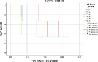
This Kaplan-Meier curve (Figure 1) shows time to tumor progression when comparing the levels of androgen receptor expression in luminal A patients generally; as follows:
▪ Score 3: 100% of patients survived for → 34 months… 64% of patients survived for Regular 57 months
▪ Score 5: 100% of patients survived for → 13 months… 75% of patients survived for → 30 months… 50% of patients survived for → 80 months
▪ Score 0: 100% → 18 months.
(with overall maximal standard error: 0.750)
With body mass index:
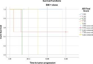
This Kaplan-Meier curve (Figure 2) shows time to tumor progression when comparing the levels of androgen receptor expression in luminal A obese patients as calculated by body mass index (BMI); as follows:
▪ Score 3: 100% → 55 months
▪ Score 8: 100% → 30 months
▪ Score 5: 100% → 14 months… 50% → 60 months
▪ Score 7: 100% → 7 months… 50% → 63 months. (with overall maximal standard error: 0.500)
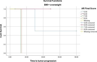
This Kaplan-Meier curve (Figure 3) shows time to tumor progression when comparing the levels of androgen receptor expression in luminal A overweight patients as calculated by Body Mass Index (BMI); as follows:
▪ Score 6: 100% → 50 months
▪ Score 5: 100% → 30 months… 50% → 80 months
▪ Score 8: 100% → 16 months
▪ Score 7: 100% → 15 months. (with overall maximal standard error: 0.500)
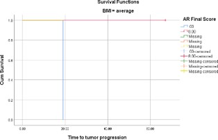
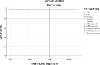
This Kaplan-Meier curve (Figure 4) shows time to tumor progression when comparing the levels of androgen receptor expression in luminal A average Body Mass Index (BMI) patients; as follows:
▪ Score 8: 100% → 70 months
▪ Score 0: 100% → 18 months. (with overall maximal standard error: 0.000)
This Kaplan-Meier curve (Figure 5) shows time to tumor progression when comparing the levels of androgen receptor expression in luminal A underweight patients as calculated by body mass index (BMI); as follows:
▪ Score 3: 100% → 34 months… 50% → 64 months
▪ Score 6: 100% → 44 months. (with overall maximal standard error: 0.500)
With age group
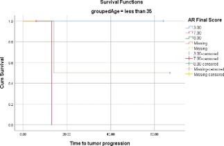
This Kaplan-Meier curve (Figure 6) shows time to tumor progression when comparing the levels of androgen receptor expression in luminal A patients with regard to age less than 35 years; as follows:
▪ Score 3: 100% → 64 months
▪ Score 8: 100% → 16 months… 50% → 68 months
▪ Score 7: 100% → 14 months.
(with overall maximal standard error: 0.500)
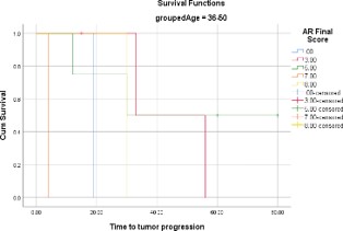
This KaplanMeier curve (Figure 7) shows time to tumor progression when comparing the levels of androgen receptor expression in luminal A patients with regard to age 36 to 50 years; as follows:
▪ Score 3: 100% → 34 months… 50% → 55 months
▪ Score 5: 100% → 12 months… 95% → 30 months… 50% → 8 months
▪ Score 8: 100% → 30 months.
(with overall maximal standard error: 0.750)
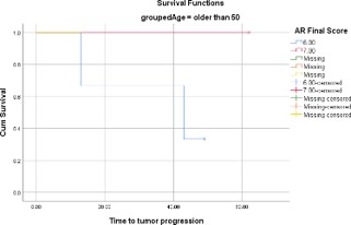
This Kaplan-Meier curve (Figure 8) shows time to tumor progression when comparing the levels of androgen receptor expression in luminal A patients with regard to age older than 50 years; as follows:
▪ Score 7: 100% → 63 months
▪ Score 6: 100% → 14 months… 66% → 43 months… 33% → 50 months. (with overall maximal standard error: 0.667)
With menopausal status
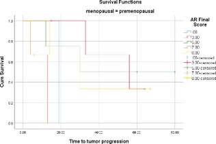
This Kaplan-Meier curve (Figure 9) shows time to tumor progression when comparing the levels of androgen receptor expression in luminal A premenopausal patients; as follows:
▪ Score 3: 100% → 33 months… 68% → 16 months
▪ Score 5: 100% → 16 months… 75% → 30 months… 50% → 80 months
▪ Score 0: 100% → 18 months. (with overall maximal standard error: 0.750)
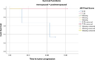
This Kaplan-Meier curve (Figure 10) shows time to tumor progression when comparing the levels of androgen receptor expression in luminal A postmenopausal patients as follows:
▪ Score 7: 100% → 62 months
▪ Score 6: 100% → 13 months… 65% → 43 months… 35% → 48 months. (with overall maximal standard error: 0.667)
With stage early versus advanced
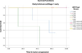
This Kaplan-Meier curve (Figure 11) shows time to tumor progression when comparing the levels of androgen receptor expression in luminal A patients with regard to early stage; as follows:
▪ Score 5: 100% → 80 months
▪ Score 8: 100% → 68 months
▪ Score 7: 100% → 62 months
▪ Score 3: 100% → 32 months… 64% → 55 months
▪ Score 6: 100% → 14 months… 50% → 44 months. (with overall maximal standard error: 0.667)
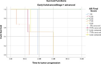
This Kaplan-Meier curve (Figure 12) shows time to tumor progression when comparing the levels of androgen receptor expression in luminal A patients with regard to advanced stage; as follows:
▪ Score 6: 100% → 48 months
▪ Score 0: 100% → 18 months
▪ Score 7: 100% → 7 months… 68% → 16 months… 50% → 30 months. (with overall maximal standard error: 0.667)
Response to Tamoxifen
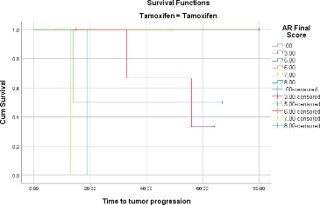
This Kaplan-Meier curve (Figure 13) shows time to tumor progression when comparing the levels of androgen receptor expression in luminal A patients with regard to response to tamoxifen; as follows:
▪ Score 5: 100% → 80 months
▪ Score 3: 100% → 34 months… 65% → 55 months… 33% →66 months
▪ Score 8: 100% → 15 months… 50 % → 68 months
▪ Score 7: 100% → 14 months… (most resistant). (with overall maximal standard error: 0.667)
Response to Letrozole
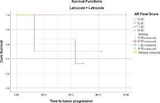
This Kaplan-Meier curve (Figure 14) shows time to tumor progression when comparing the levels of androgen receptor expression in luminal A patients with regard to response to letrozole; as follows:
▪ Score 5: 100% → 80 months
▪ Score 7: 100% → 13 months… 50% → 64 months
Score 8: 100% → 30 months. (with overall maximal standard error: 0.667)
Summary of TTP/PFS curves in luminal A Subgroup
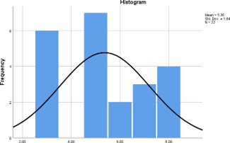
This histogram (Figure 15) shows that score 5 as degree of androgen receptor expression is the predominant and has the best Time to Tumor Progression (TTP) in all luminal A patients in our study. Z score = -7.386. P-Value is < .00001. (statistically significant)
Overall survival:
Generally:
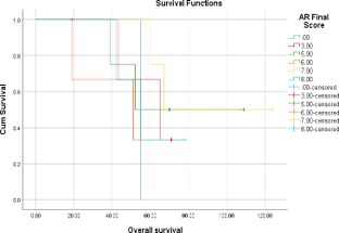
This Kaplan-Meier curve (Figure 16) shows the overall survival when comparing the levels of androgen receptor expression in luminal A patients generally; as follows:
▪ Score 7: 100% of patients survived for → 60 months… 78% of patients survived for → 68 months… 50% survived for → 125 months
▪ Score 5: 100% → 38 months… 74% → 55 months… 50% → 110 months
▪ Score 0: 100% → 55 months (with maximal standard error: 0.272)
With Body Mass Index:
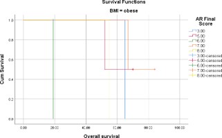
This Kaplan-Meier curve (Figure 17) shows the overall survival when comparing the levels of androgen receptor expression in luminal A obese patients as estimated by Body mass Index (BMI); as follows:
▪ Score 7: 100% → 66 months… 50% → 85 months
▪ Score 3: 100% → 64 months
▪ Score 8: 100% → 55 months
Score 5: 100% → 50 months… 50% → 70 months (with maximal standard error: 0.354)
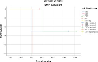
This Kaplan-Meier curve (Figure 18) shows the overall survival when comparing the levels of androgen receptor expression in luminal A overweight patients as estimated by Body mass Index (BMI); as follows:
▪ Score 7: 100% → 60 months… 50% → 125 months
▪ Score 5: 100% → 48 months… 50% → 110 months
▪ Score 6: 100% → 55 months
▪ Score 8: 100% → 43 months (with maximal standard error: 0.354)
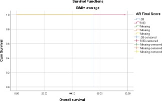
This Kaplan-Meier curve (Figure 19) shows the overall survival when comparing the levels of androgen receptor expression in luminal A patients with average Body Mass Index (BMI); as follows:
▪ Score 8: 100% → 78 months
▪ Score 0: 100% → 55 months (with maximal standard error: 0.000)
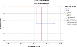
This Kaplan-Meier curve (Figure 20) shows the overall survival in luminal A underweight patients, as estimated by the Body Mass Index (BMI) as follows:
▪ Score 6: 100% → 52 months
▪ Score 3: 100% → 52 months … 50% → 72 months. (with maximal standard error: 0.354)
With age group:
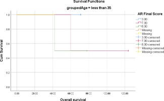
This Kaplan-Meier curve (Figure 21) shows the overall survival in luminal A patients aging less than 35 years; as follows:
▪ Score 7: 100% → 60 months… 50% → 125 months
▪ Score 3: 100% → 72 months
▪ Score 8: 100% → 42 months… 50% → 78 months.(with maximal standard error: 0.354)
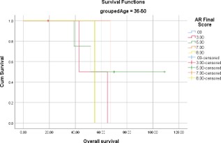
This Kaplan-Meier curve (Figure 22) shows the overall survival in luminal A patients aging 36 to 50 years; as follows:
Score 7: 100% → 68 months
Score 5: 100% → 39 months… 75 % → 52 months… 50% →110 months
Score 8: 100% → 55 months
Score 3: 100% → 44 months… 50% → 66 months.(with maximal standard error: 0.354)
Score 5: 100% → 38 months… 75% → 54 months… 50% → 110 months
Score 8: 100% → 42 months… 68% → 56 months… 36% → 78 months (with maximal standard error: 0.272)
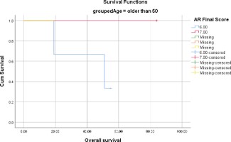
This Kaplan-Meier curve (Figure 23) shows the overall survival in luminal A patients aging older than 50 years; as follows:
▪ Score 7: 100% → 85 months
▪ Score 6: 100% → 19 months… 68 % → 52 months… 36% → 55 months (with maximal standard error: 0.272)
With menopausal
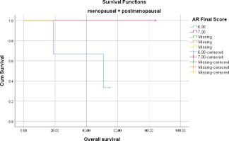
This Kaplan-Meier curve (Figure 24) shows the overall survival in luminal A postmenopausal patients; as follows:
▪ Score 7: 100% → 85 months
▪ Score 6: 100% → 18 months… 68% → 52 months… 34% → 55 months (with maximal standard error: 0.272) With stage early versus advanced
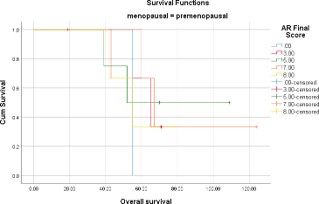
This Kaplan-Meier curve (Figure 25) shows the overall survival in luminal A premenopausal patients; as follows:
▪ Score 7: 100% → 60 months… 66% → 68 months… 33% → 125 months
▪ Score 0: 100% → 56 months
▪ Score 3: 100% → 44 months… 68 % → 68 months… 35% → 70 months
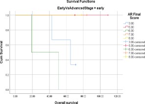
This Kaplan-Meier curve (Figure 26) shows the overall survival in luminal A patients with early stage; as follows:
▪ Score 5: 100% → 110 months
▪ Score 8: 100% → 78 months
▪ Score 3: 100% → 44 months… 68 % → 68 months… 35% → 70 months
▪ Score 6: 100% → 19 months… 50% → 52 months (with maximal standard error: 0.354)
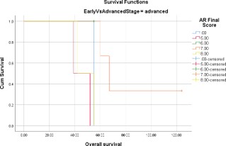
This Kaplan-Meier curve (Figure 27) shows the overall survival in luminal A patients with advanced stage; as follows:
▪ Score 7: 100% → 60 months… 66% → 68 months… 33% → 125 months
▪ Score 0: 100% → 56 months
▪ Score 8: 100% → 42 months… 50 % → 68 months
▪ Score 5: 100% → 38 months… 50% → 60 months (with maximal standard error: 0.354)
Summary of OS curved of luminal A subgroup
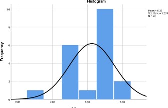
This histogram (Figure 28) shows that score 5 as degree of androgen receptor expression is the predominant and has the Overall Survival (OS) in all luminal A patients in our study. Z score =-17.96. P-Value is <.00001. (statistically significant)
Discussion
◆ Vera-Badillo FE in 2014 found -in a large systematic review that included 7693 breast cancers among 19 studiesthat AR expression was 74.8% in ER positive tumors and 31.8% in ER negative tumors [17].
◆ AR was shown to be expressed up to 90% of ERα positive BC [5].
▸ Here, in our study, 50 (96.1%) out of all our 52 hormonal receptor positive patients stained positive for androgen receptor.
✚ Our results came in concordance with was stated by Giovenalli P and in contradiction with what was stated by Vera-Badillo FE.
◆ Hickey TE and colleagues had published in 2012 that AR might be a potential tumor suppressor in ER-positive breast cancer, with anti-proliferative effects due to cross talk between the steroid receptor signaling pathways [8].
◆ Cochrane and colleagues had demonstrated inhibition of both dihydrotestosterone-mediated and estradiol-mediated proliferation of ERpositive and AR-positive breast cancer cells by antiandrogens [1].
▸ In our study, intermediate androgen receptor expression (score 5) had the best time to both tumor progression and overall survival in all hormonal receptor positive. (P-Value <0.00001) (statistically significant)
✚ Our study results came in agreement with what was stated by Hickey TE, Cochrane and their colleagues.
◆ In 2014, Rechoum Y studied ER-positive breast cancer cells in vitro to investigate variable molecules and the efficacy of variable treatment options. His study showed that: In ER-positive MCF-7 cells engineered to overexpress aromatase and AR (MCF-7 AR Arom cells) treated with Androstenedione (AD), the steroidal AI, anastrozole (Ana) does not inhibit growth and ER transcriptional activity. This effect could be due to an increase in pAKT (phosphorylated AKT, AKT is an oncogene; also known as protein kinase B “PKB”) and pIGF-1R levels as well as to a co-operation between AR and ER that might induce the up-regulation of AR and ER-responsive gene expression. AR antagonists, or the antiestrogen fulvestrant or the IGF-1R and AKT inhibitors restore the sensitivity to Anastrozole [14].
◆ He also concluded that AR is also responsible for the Aromatase Inhibitor (AI) resistance in ER-positive BC cells [14].
▸ In our study, Letrozole effect has been shown to be better with the intermediate androgen receptor expression followed by the high levels of androgen receptor expression.
✚ As both are reversible aromatase inhibitors, this finding comes in concordance with what was found by Rechoum Y.
◆ In December 2020, Krob I and the investigating team conducted a randomized Placebo Controlled Phase II Trial Evaluating Exemestane with or without Enzalutamide in Patients with Hormone Receptor–Positive Breast Cancer and concluded that PFS was not improved by the addition of enzalutamide to exemestane in hormone receptor positive androgen receptor positive breast cancer patients [10].
▸ In our study, Letrozole effect has been shown to be better with the intermediate androgen receptor expression followed by the high levels of androgen receptor expression.
✚ This finding came in contradiction with what was found by Krob I.
◆ Kevin H. Kensler and his colleagues stated that an AR:ER ratio ≥2 was associated with an increased rate of tamoxifen failure in a cohort of 192 women with ER+ breast cancer [9].
▸ In our study, among breast cancer female patients who received Tamoxifen, positive androgen receptor negatively affected their response to Tamoxifen. The high levels of expression of androgen receptor had the worst response to Tamoxifen. The intermediate degree of expression of androgen receptor had the best prognosis.
✚ Our study results came in concordance with what was stated by Kevin H. Kensler and his colleagues.
Conclusion
Androgen receptor intermediate expression had the best prognosis in luminal A, invasive breast cancer female patients.
Concerning breast cancer female patients who received Tamoxifen, positive androgen receptor negatively affected their response to Tamoxifen. The high levels of expression of androgen receptor had the worst response to Tamoxifen. The intermediate degree of expression of androgen receptor had the best prognosis.
Accidentally, Letrozole effect had been shown to be better with the intermediate androgen receptor expression followed by the high levels of androgen receptor expression. The intermediately positive androgen receptor breast cancer female patients had best response when compared to the highly positive androgen receptor ones.
References
- Cochrane DR, Bernales S, et al. Role of the androgen receptor in breast cancer and preclinical analysis of enzalutamide. Breast Cancer Res. 2014; 16: R7.
- De Amicis F, Thirugnansampanthan J, Cui Y, Selever J, Beyer A, Parra I, et al. Androgen receptor overexpression induces tamoxifen resistance in human breast cancer cells. Breast Cancer Res Treat. 2010; 121: 1-11.
- Hilborn E, Gacic J, Fornander T, et al. Androgen receptor expression predicts beneficial tamoxifen response in oestrogen receptorα-negative breast cancer. Br J Cancer. 2016; 114: 248-255.
- De Amicis F, Thirugnansampanthan J, Cui Y, et al. Androgen receptor overexpression induces tamoxifen resistance in human breast cancer cells. Breast Cancer Res Treat. 2010; 121: 1-11.
- Giovanelli P, et al. The androgen receptor in breast cancer. Front Endocrinol. 2018; 9: 492.
- Gucalp A, et al. Phase II trial of bicalutamide in patients with androgen receptor-positive, estrogen receptor-negative metastatic breast cancer. Clin Cancer Res. 2013; 19: 5505-5512.
- Helwick C. Targeting the androgen receptor in breast cancer. ASCO Post. 2017.
- Hickey TE, et al. The androgen receptor in breast tissues: growth inhibitor, tumor suppressor, oncogene? Mol Endocrinol. 2012; 26: 1252-1267.
- Kensler KH, et al. Prognostic and predictive value of androgen receptor expression in postmenopausal women with estrogen receptor-positive breast cancer: results from the Breast International Group Trial 1–98. Breast Cancer Res. 2019; 21: 42.
- Krob I, et al. A randomized placebo-controlled phase II trial evaluating exemestane with or without enzalutamide in patients with hormone receptor-positive breast cancer. Clin Cancer Res. 2020; 26: 6149-6159.
- Lebert JM, et al. Advances in the systemic treatment of triple-negative breast cancer. Curr Oncol. 2018; 25: 119-128.
- Park S, et al. Expression of androgen receptors in primary breast cancer. Ann Oncol. 2010; 21: 488-492.
- Raosoft Inc. Sample size calculator. Raosoft. 2004.
- Rechoum Y, Rovito D, Iacopetta D, Barone I, Andò S, Weigel NL, et al. AR collaborates with ER-alpha in aromatase inhibitor-resistant breast cancer. Breast Cancer Res Treat. 2014; 147: 473-485.
- Rizza P, Barone I, Zito D, Giordano F, Lanzino M, De Amicis F, et al. Estrogen receptor beta as a novel target of androgen receptor action in breast cancer cell lines. Breast Cancer Res. 2014; 16: R21.
- Suez Canal University Clinical Oncology Department. Confidential archive files. Ismailia: Suez Canal University; 2008–2022.
- Vera-Badillo FE, et al. Androgen receptor expression and outcomes in early breast cancer: a systematic review and meta-analysis. J Natl Cancer Inst. 2014; 106: djt319.